The decision to treat a rotator cuff tear surgically or non-surgically should be based on several factors, including size of tear, patient age, expected activity level, degree of tendon retraction, and presence of rotator cuff muscle atrophy and fatty replacement.
Earlier surgical intervention may be needed when there is weakness and substantial functional disability, or if pain continues despite several months of physiotherapy and medical therapy.[32]Oh LS, Wolf BR, Hall MP, et al. Indications for rotator cuff repair: a systematic review. Clin Orthop Relat Res. 2007 Feb;455:52-63.
http://www.ncbi.nlm.nih.gov/pubmed/17179786?tool=bestpractice.com
[33]Seida JC, LeBlanc C, Schouten JR, et al. Systematic review: nonoperative and operative treatments for rotator cuff tears. Ann Intern Med. 2010 Aug 17;153(4):246-55.
http://www.ncbi.nlm.nih.gov/pubmed/20621893?tool=bestpractice.com
[34]Agency for Healthcare Research and Quality (US). Comparative effectiveness of nonoperative and operative treatments for rotator cuff tears. July 2010 [internet publication].
https://www.ncbi.nlm.nih.gov/books/NBK47305
http://www.ncbi.nlm.nih.gov/pubmed/21028756?tool=bestpractice.com
In addition, if the patient requires full use of the arm for vocational or recreational needs, then repair should be considered.
The time since injury is also an important consideration, as the function and appearance of a torn rotator cuff generally deteriorate with time. With chronic tears, muscle tissue may atrophy in time and is replaced with fatty tissue, often termed fatty degeneration.[31]Melis B, DeFranco MJ, Chuinard C, et al. Natural history of fatty infiltration and atrophy of the supraspinatus muscle in rotator cuff tears. Clin Orthop Relat Res. 2010 Jun;468(6):1498-505.
https://pmc.ncbi.nlm.nih.gov/articles/PMC2865597
http://www.ncbi.nlm.nih.gov/pubmed/20094853?tool=bestpractice.com
Risk factors for re-tear after arthroscopic (minimally invasive) rotator cuff repair include fatty infiltration (subscapularis and infraspinatus) and symptom duration.[35]Zhao J, Luo M, Pan J, et al. Risk factors affecting rotator cuff retear after arthroscopic repair: a meta-analysis and systematic review. J Shoulder Elbow Surg. 2021 Nov;30(11):2660-70.
https://www.jshoulderelbow.org/article/S1058-2746(21)00477-8/fulltext
http://www.ncbi.nlm.nih.gov/pubmed/34089878?tool=bestpractice.com
A direct correlation has been described between the extent of the fatty degeneration of the rotator cuff muscle and the time from injury.[36]Goutallier D, Postel JM, Bernageau J, et al. Fatty muscle degeneration in cuff ruptures: pre- and postoperative evaluation by CT scan. Clin Orthop. 1994 Jul;(304):78-83.
http://www.ncbi.nlm.nih.gov/pubmed/8020238?tool=bestpractice.com
Researchers found improved outcomes and a reduced re-tear rate when repair was performed when fatty degeneration was minimal. In patients over 60 years of age, a favourable outcome can still be expected after repair, provided tissue quality remains sufficient.[37]Downie BK, Miller BS. Treatment of rotator cuff tears in older individuals: a systematic review. J Shoulder Elbow Surg. 2012 Sep;21(9):1255-61.
http://www.ncbi.nlm.nih.gov/pubmed/22365558?tool=bestpractice.com
Acute tears (identified within 6 weeks of a significant known trauma)
Treatment options for acute tears are determined in large part by the size of tear and how symptomatic the patient is at the time of presentation. Involvement of a physiotherapist is often helpful. If there is external rotation weakness, then infraspinatus involvement is present and surgery is more likely to be necessary since shoulder biomechanics are adversely affected in the presence of two tendon tears.
Surgical repair of acute small tears
Surgical repair is the first-line treatment for patients with good functional status, especially if functional demands are high. When surgery is undertaken before the onset of atrophy, more predictable results are ensured. One randomised controlled trial of people with rotator cuff tear not exceeding 3 cm found that primary tendon repair improved pain, motion, and strength at 10 years compared with physiotherapy.[38]Moosmayer S, Lund G, Seljom US, et al. At a 10-year follow-up, tendon repair is superior to physiotherapy in the treatment of small and medium-sized rotator cuff tears. J Bone Joint Surg Am. 2019 Jun 19;101(12):1050-60.
http://www.ncbi.nlm.nih.gov/pubmed/31220021?tool=bestpractice.com
Surgical options include arthroscopic, mini-open, and open repair. The primary goal is to provide a pain-free joint with good function.
Conservative measures for small acute tears
Non-surgical options should be considered first for older and sedentary patients with small tears with mild loss of range of motion (ROM) and strength, and for patients with low functional demands. Ice, stretching, and non-steroidal anti-inflammatory drugs (NSAIDs) are the initial treatments. Once ROM returns (usually at about 4 weeks), toning exercises can be started while stretching continues. A single subacromial corticosteroid injection can be used to control inflammation and reduce pain if rehabilitation therapy and NSAIDs are ineffective.[39]Weber S, Chahal J. Management of rotator cuff injuries. J Am Acad Orthop Surg. 2020 Mar 1;28(5):e193-201.
https://journals.lww.com/jaaos/Fulltext/2020/03010/Management_of_Rotator_Cuff_Injuries.4.aspx
http://www.ncbi.nlm.nih.gov/pubmed/31599763?tool=bestpractice.com
Rehabilitation can resume a few days after the injection.[40]Coombes BK, Bisset L, Vicenzino B. Efficacy and safety of corticosteroid injections and other injections for management of tendinopathy: a systematic review of randomised controlled trials. Lancet. 2010 Nov 20;376(9754):1751-67.
http://www.ncbi.nlm.nih.gov/pubmed/20970844?tool=bestpractice.com
Repeated injections are to be avoided, as healing of a potential surgical repair may be compromised.[41]Puzzitiello RN, Patel BH, Nwachukwu BU, et al. Adverse impact of corticosteroid injection on rotator cuff tendon health and repair: a systematic review. Arthroscopy. 2020 May;36(5):1468-75.
http://www.ncbi.nlm.nih.gov/pubmed/31862292?tool=bestpractice.com
Contraindications for subacromial corticosteroid injections include septic arthritis, a previous adverse reaction, or systemic infection.[41]Puzzitiello RN, Patel BH, Nwachukwu BU, et al. Adverse impact of corticosteroid injection on rotator cuff tendon health and repair: a systematic review. Arthroscopy. 2020 May;36(5):1468-75.
http://www.ncbi.nlm.nih.gov/pubmed/31862292?tool=bestpractice.com
One systematic review and meta-analysis suggested that NSAIDs are less effective than corticosteroid injection at achieving remission in patients with shoulder pain at 4-6 weeks after treatment.[42]Zheng XQ, Li K, Wei YD, et al. Nonsteroidal anti-inflammatory drugs versus corticosteroid for treatment of shoulder pain: a systematic review and meta-analysis. Arch Phys Med Rehabil. 2014 Oct;95(10):1824-31.
http://www.ncbi.nlm.nih.gov/pubmed/24841629?tool=bestpractice.com
However, as this review included a limited number of small studies, the results should be interpreted with caution.[42]Zheng XQ, Li K, Wei YD, et al. Nonsteroidal anti-inflammatory drugs versus corticosteroid for treatment of shoulder pain: a systematic review and meta-analysis. Arch Phys Med Rehabil. 2014 Oct;95(10):1824-31.
http://www.ncbi.nlm.nih.gov/pubmed/24841629?tool=bestpractice.com
Activity should be modified to reduce overhead lifting. When returns ROM (usually around 4 weeks), toning should be added while stretching is continued.
Lack of response to at least 10-12 weeks of medical and physiotherapy should prompt consideration of open, mini-open, or arthroscopic repair for patients with non-operative treatment for smaller or partial tears. Surgeons generally should perform the technique that provides the best and most reliable results in their hands.
[Figure caption and citation for the preceding image starts]: Subacromial injection. Insert needle just inferior to posterior edge of acromion (x), aiming parallel to the undersurface of the acromionFrom the collection of Daniel J. Solomon, MD; used with permission [Citation ends].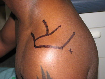
Management of acute medium, large, or massive reparable tears
Surgical repair is the first-line treatment for patients with good functional status, especially if functional demands are high , or the injury is on the dominant side. Options include arthroscopic, mini-open, and open repair. The primary goal is to provide a pain-free joint with good function.
If the patient is older and sedentary, rehabilitation therapy with NSAIDs, ice, stretching, and exercise should be considered before surgery. Rehabilitation should be managed by a physiotherapist. Focus should be on strengthening remaining cuff, deltoid, and scapular stabilisers, as well as posterior capsular stretching.
Irreparable tears
Debridement may be appropriate for patients with pain as their predominant symptom and lower demands for shoulder strength. The ideal debridement patient has good deltoid function and an intact coracoacromial arch.
Arthroscopic debridement can include bursectomy, acromioclavicular joint resection, and smoothing of the greater tuberosity. Subacromial decompression alone for the treatment of symptomatic rotator cuff disease presenting with impingement features and without full‐thickness rotator cuff tears does not provide clinically important benefits compared with placebo surgery with respect to pain, function, or quality of life, and is no longer routinely recommended.[39]Weber S, Chahal J. Management of rotator cuff injuries. J Am Acad Orthop Surg. 2020 Mar 1;28(5):e193-201.
https://journals.lww.com/jaaos/Fulltext/2020/03010/Management_of_Rotator_Cuff_Injuries.4.aspx
http://www.ncbi.nlm.nih.gov/pubmed/31599763?tool=bestpractice.com
[43]Karjalainen TV, Jain NB, Page CM, et al. Subacromial decompression surgery for rotator cuff disease. Cochrane Database Syst Rev. 2019 Jan 17;1(1):CD005619.
https://www.cochranelibrary.com/cdsr/doi/10.1002/14651858.CD005619.pub3/full
http://www.ncbi.nlm.nih.gov/pubmed/30707445?tool=bestpractice.com
[44]Vandvik PO, Lähdeoja T, Ardern C, et al. Subacromial decompression surgery for adults with shoulder pain: a clinical practice guideline. BMJ. 2019 Feb 6;364:l294.
http://www.ncbi.nlm.nih.gov/pubmed/30728120?tool=bestpractice.com
BMJ: subacromial decompression surgery for adults with shoulder pain: a clinical practice guideline
Opens in new window However, decompression in the presence of rotator cuff repair may, in some cases, lower recurrent tear rates.[45]MacDonald P, McRae S, Leiter J, et al. Arthroscopic rotator cuff repair with and without acromioplasty in the treatment of full-thickness rotator cuff tears: a multicenter, randomized controlled trial. J Bone Joint Surg Am. 2011 Nov 2;93(21):1953-60.
http://www.ncbi.nlm.nih.gov/pubmed/22048089?tool=bestpractice.com
Reverse shoulder arthroplasty (RSA) is an established surgical option that is used for the treatment of massive, irreparable rotator cuff tear with or without arthritis in cuff tear arthropathy settings, or in elderly patients with dislocation and cuff tear that fail non-operative management, or rotator cuff tear.[46]St Pierre P. When is a reverse shoulder arthroplasty indicated for a rotator cuff tear? Sports Med Arthrosc Rev. 2024 Mar 1;32(1):17-21.
http://www.ncbi.nlm.nih.gov/pubmed/38695499?tool=bestpractice.com
[47]Di Benedetto P, Mancuso F, Tosolini L, et al. Treatment options for massive rotator cuff tears: a narrative review. Acta Biomed. 2021 Jul 26;92(s3):e2021026.
https://pmc.ncbi.nlm.nih.gov/articles/PMC8420830
http://www.ncbi.nlm.nih.gov/pubmed/34313657?tool=bestpractice.com
Systematic reviews and meta-analyses indicate that newer designed prostheses that offer a lateralised centre of rotation RSA may be the preferred approach.[48]Berton A, Gulotta LV, Longo UG, et al. Medialized versus lateralized center of rotation in reverse total shoulder arthroplasty: a systematic review and meta-analysis. J Clin Med. 2021 Dec 14;10(24):5868.
https://www.ncbi.nlm.nih.gov/pmc/articles/PMC8703399
http://www.ncbi.nlm.nih.gov/pubmed/34945160?tool=bestpractice.com
[49]Samitier G, Alentorn-Geli E, Torrens C, et al. Reverse shoulder arthroplasty. Part 1: systematic review of clinical and functional outcomes. Int J Shoulder Surg. 2015 Jan-Mar;9(1):24-31.
https://www.ncbi.nlm.nih.gov/pmc/articles/PMC4325387
http://www.ncbi.nlm.nih.gov/pubmed/25709242?tool=bestpractice.com
Tendon transfer is appropriate for patients with high demands for shoulder strength. Patients must be able and willing to perform extensive postoperative rehabilitation. Muscle transfers have been effective for anterosuperior tears involving the subscapularis and supraspinatus, and for posterosuperior tears involving the supraspinatus and infraspinatus. Effective donors are the pectoralis major for anterosuperior tears and the latissimus dorsi for posterosuperior tear.[50]Warner JJ. Management of massive irreparable rotator cuff tears: the role of tendon transfer. Instr Course Lect. 2001 Feb;50(6):63-71.
http://www.ncbi.nlm.nih.gov/pubmed/11372361?tool=bestpractice.com
[51]Gerber C, Hersche O. Tendon transfers for the treatment of irreparable rotator cuff defects. Orthop Clin North Am. 1997 Apr;28(2):195-203.
http://www.ncbi.nlm.nih.gov/pubmed/9113715?tool=bestpractice.com
Lower trapezius transfer is becoming increasingly popular, because this transfer closely mimics the line of pull of the infraspinatus.[52]Wagner ER, Elhassan BT. Surgical management of massive irreparable posterosuperior rotator cuff tears: arthroscopic-assisted lower trapezius transfer. Curr Rev Musculoskelet Med. 2020 Oct;13(5):592-604.
https://www.ncbi.nlm.nih.gov/pmc/articles/PMC7474727
http://www.ncbi.nlm.nih.gov/pubmed/32661919?tool=bestpractice.com
[53]Elhassan BT, Wagner ER, Werthel JD. Outcome of lower trapezius transfer to reconstruct massive irreparable posterior-superior rotator cuff tear. J Shoulder Elbow Surg. 2016 Aug;25(8):1346-53.
http://www.ncbi.nlm.nih.gov/pubmed/26968088?tool=bestpractice.com
Superior capsular reconstruction prevents superior humeral head migration, resulting in enhanced deltoid function. This procedure may be offered to patients with isolated irreparable rupture of the supraspinatus, where medical treatment has not been successful.[54]Werthel JD, Vigan M, Schoch B, et al. Superior capsular reconstruction - a systematic review and meta-analysis. Orthop Traumatol Surg Res. 2021 Dec;107(8s):103072.
https://www.sciencedirect.com/science/article/pii/S1877056821003170
http://www.ncbi.nlm.nih.gov/pubmed/34560311?tool=bestpractice.com
Other indications include a massive, irreparable tear of the rotator cuff or an intact or repairable subscapularies tendon (if there is intolerable pain despite conservative treatment and minimal or absent evidence of arthritis), or for patients who do not wish to undergo arthroplasty.[54]Werthel JD, Vigan M, Schoch B, et al. Superior capsular reconstruction - a systematic review and meta-analysis. Orthop Traumatol Surg Res. 2021 Dec;107(8s):103072.
https://www.sciencedirect.com/science/article/pii/S1877056821003170
http://www.ncbi.nlm.nih.gov/pubmed/34560311?tool=bestpractice.com
[55]Claro R, Fonte H. Superior capsular reconstruction: current evidence and limits. EFORT Open Rev. 2023 May 9;8(5):340-50.
https://pmc.ncbi.nlm.nih.gov/articles/PMC10233801
http://www.ncbi.nlm.nih.gov/pubmed/37158430?tool=bestpractice.com
[56]Mihata T, Lee TQ, Watanabe C, et al. Clinical results of arthroscopic superior capsule reconstruction for irreparable rotator cuff tears. Arthroscopy. 2013 Mar;29(3):459-70.
https://www.arthroscopyjournal.org/article/S0749-8063(12)01789-6/fulltext
http://www.ncbi.nlm.nih.gov/pubmed/23369443?tool=bestpractice.com
The results have been inconsistent but investigators maintain that technique success depends on graft type and thickness.[54]Werthel JD, Vigan M, Schoch B, et al. Superior capsular reconstruction - a systematic review and meta-analysis. Orthop Traumatol Surg Res. 2021 Dec;107(8s):103072.
https://www.sciencedirect.com/science/article/pii/S1877056821003170
http://www.ncbi.nlm.nih.gov/pubmed/34560311?tool=bestpractice.com
[55]Claro R, Fonte H. Superior capsular reconstruction: current evidence and limits. EFORT Open Rev. 2023 May 9;8(5):340-50.
https://pmc.ncbi.nlm.nih.gov/articles/PMC10233801
http://www.ncbi.nlm.nih.gov/pubmed/37158430?tool=bestpractice.com
Chronic tears
In general, chronic tears should be treated initially with conservative therapies (e.g., ice and stretching, cuff strengthening, scapular stabilisation, NSAIDs, and subacromial corticosteroid injections). Involvement of a physiotherapist is often helpful.
Surgery can be pursued if a tear is unresponsive to non-operative treatment after 10-12 weeks. Younger patients are typically treated with a more aggressive approach, with surgery considered earlier in their treatment course, especially if they complain of weakness.[32]Oh LS, Wolf BR, Hall MP, et al. Indications for rotator cuff repair: a systematic review. Clin Orthop Relat Res. 2007 Feb;455:52-63.
http://www.ncbi.nlm.nih.gov/pubmed/17179786?tool=bestpractice.com
Patients must be engaged in their treatment. The patient’s belief in the success of rehabilitation is the strongest predictor of non-operative outcome, more so than tear size.[57]Dunn WR, Kuhn JE, Sanders R, et al. 2013 Neer Award: predictors of failure of nonoperative treatment of chronic, symptomatic, full-thickness rotator cuff tears. J Shoulder Elbow Surg. 2016 Aug;25(8):1303-11.
http://www.ncbi.nlm.nih.gov/pubmed/27422460?tool=bestpractice.com
In an older patient with a large chronic tear, the quality of the tissue is often less than optimal for healing. These patients and others with low functional demands are frequently less interested in powerful overhead actions and more interested in pain relief with a functional arc of motion. A well-designed, non-operative rehabilitation programme, consisting of stretching and strengthening, can often attain these goals.[57]Dunn WR, Kuhn JE, Sanders R, et al. 2013 Neer Award: predictors of failure of nonoperative treatment of chronic, symptomatic, full-thickness rotator cuff tears. J Shoulder Elbow Surg. 2016 Aug;25(8):1303-11.
http://www.ncbi.nlm.nih.gov/pubmed/27422460?tool=bestpractice.com
[58]Williams GR Jr, Rockwood CA Jr, Bigliani LU, et al. Rotator cuff tears: why do we repair them? J Bone Joint Surg. 2004 Dec;86(12):2764-76
http://www.ncbi.nlm.nih.gov/pubmed/15590865?tool=bestpractice.com
The focus of this rehabilitation is pain control, restoration of full passive motion, and optimisation of rotator cuff and periscapular muscle strength and co-ordination.
Subacromial corticosteroid injection can be used if symptoms limit rehabilitation exercises, but should be used with caution if surgery is a reasonable possibility due to the adverse effects of corticosteroids on cuff healing and infection risk.[41]Puzzitiello RN, Patel BH, Nwachukwu BU, et al. Adverse impact of corticosteroid injection on rotator cuff tendon health and repair: a systematic review. Arthroscopy. 2020 May;36(5):1468-75.
http://www.ncbi.nlm.nih.gov/pubmed/31862292?tool=bestpractice.com
Exercises can be resumed a few days after injection.
One meta-analysis found that suprascapular nerve block had similar efficacy compared with intra-articular corticosteroid injection for shoulder pain, and may be used as an adjunct therapy if corticosteroid injection alone does not provide sustained pain relief.[59]Chang KV, Hung CY, Wu WT, et al. Comparison of the effectiveness of suprascapular nerve block with physical therapy, placebo, and intra-articular injection in management of chronic shoulder pain: a meta-analysis of randomized controlled trials. Arch Phys Med Rehabil. 2016 Aug;97(8):1366-80.
http://www.ncbi.nlm.nih.gov/pubmed/26701762?tool=bestpractice.com
For patients with considerable pain after 10-12 weeks of therapy, the following surgical options should be considered on a case-by-case basis.[32]Oh LS, Wolf BR, Hall MP, et al. Indications for rotator cuff repair: a systematic review. Clin Orthop Relat Res. 2007 Feb;455:52-63.
http://www.ncbi.nlm.nih.gov/pubmed/17179786?tool=bestpractice.com
[50]Warner JJ. Management of massive irreparable rotator cuff tears: the role of tendon transfer. Instr Course Lect. 2001 Feb;50(6):63-71.
http://www.ncbi.nlm.nih.gov/pubmed/11372361?tool=bestpractice.com
[51]Gerber C, Hersche O. Tendon transfers for the treatment of irreparable rotator cuff defects. Orthop Clin North Am. 1997 Apr;28(2):195-203.
http://www.ncbi.nlm.nih.gov/pubmed/9113715?tool=bestpractice.com
[52]Wagner ER, Elhassan BT. Surgical management of massive irreparable posterosuperior rotator cuff tears: arthroscopic-assisted lower trapezius transfer. Curr Rev Musculoskelet Med. 2020 Oct;13(5):592-604.
https://www.ncbi.nlm.nih.gov/pmc/articles/PMC7474727
http://www.ncbi.nlm.nih.gov/pubmed/32661919?tool=bestpractice.com
[53]Elhassan BT, Wagner ER, Werthel JD. Outcome of lower trapezius transfer to reconstruct massive irreparable posterior-superior rotator cuff tear. J Shoulder Elbow Surg. 2016 Aug;25(8):1346-53.
http://www.ncbi.nlm.nih.gov/pubmed/26968088?tool=bestpractice.com
[56]Mihata T, Lee TQ, Watanabe C, et al. Clinical results of arthroscopic superior capsule reconstruction for irreparable rotator cuff tears. Arthroscopy. 2013 Mar;29(3):459-70.
https://www.arthroscopyjournal.org/article/S0749-8063(12)01789-6/fulltext
http://www.ncbi.nlm.nih.gov/pubmed/23369443?tool=bestpractice.com
[60]Beaudreuil J, Dhénain M, Coudane H, Mlika-Cabanne N. Clinical practice guidelines for the surgical management of rotator cuff tears in adults. Orthop Traumatol Surg Res. 2010 Apr;96(2):175-9.
http://www.ncbi.nlm.nih.gov/pubmed/20464793?tool=bestpractice.com
[61]Izquierdo R, Voloshin I, Edwards S, et al; American Academy of Orthopedic Surgeons. Treatment of glenohumeral osteoarthritis. J Am Acad Orthop Surg. 2010 Jun;18(6):375-82.
http://www.ncbi.nlm.nih.gov/pubmed/20511443?tool=bestpractice.com
[62]Stewart RK, Kaplin L, Parada SA, et al. Outcomes of subacromial balloon spacer implantation for massive and irreparable rotator cuff tears: a systematic review. Orthop J Sports Med. 2019 Oct;7(10):2325967119875717.
https://www.ncbi.nlm.nih.gov/pmc/articles/PMC6794659
http://www.ncbi.nlm.nih.gov/pubmed/31663007?tool=bestpractice.com
Arthroscopic, mini-open, or open surgical repair: typically considered for patients with both pain and functional limitations who are anticipating return to an active lifestyle.
Debridement: typically used for patients with minimal functional limitations but pain as a primary complaint, and for patients with limited functional goals and expectations. Tuberoplasty can be performed when there is a prominent greater tuberosity, which may abut the acromion on abduction.
Hemiarthroplasty and reverse total shoulder arthroplasty: salvage procedures for patients who have long-standing tears and develop cuff tear arthropathy.
Superior capsule reconstruction: typically used for younger active patients with irreparable tears. The procedure employs a graft to reconstruct the superior capsule and may inhibit proximal humeral migration. This technique should not be used in the presence of advanced arthropathy or irreparable subscapularis tear.
Balloon spacer: indicated for irreparable tears in patients who are not suitable candidates for arthroplasty. An inflatable biodegradable balloon is inserted under the acromion to act as a physical barrier to reduce subacromial friction. This device may help enhance deltoid rehabilitation; however, the reported success of this intervention has been inconsistent.[63]Sirignano M, Nyland J, Krupp R. Subacromial balloon spacer massive rotator cuff tear treatment systematic review and meta-analysis: patient selection and physical therapy may be keys to outcome success. Knee Surg Sports Traumatol Arthrosc. 2024 Sep;32(9):2346-57.
http://www.ncbi.nlm.nih.gov/pubmed/38922784?tool=bestpractice.com
[64]Singh S, Reeves J, Langohr GDG, et al. The effect of the subacromial balloon spacer on humeral head translation in the treatment of massive, irreparable rotator cuff tears: a biomechanical assessment. J Shoulder Elbow Surg. 2019 Oct;28(10):1841-7.
http://www.ncbi.nlm.nih.gov/pubmed/31272890?tool=bestpractice.com
[65]Liu F, Dong J, Kang Q, et al. Subacromial balloon spacer implantation for patients with massive irreparable rotator cuff tears achieves satisfactory clinical outcomes in the short and middle of follow-up period: a meta-analysis. Knee Surg Sports Traumatol Arthrosc. 2021 Jan;29(1):143-53.
http://www.ncbi.nlm.nih.gov/pubmed/31894368?tool=bestpractice.com
A balloon may also be used to ‘protect’ a cuff repair as well as prevent subacromial adhesions.[66]Garofalo R, De Crescenzo A, Fontanarosa A, et al. Rotator cuff repair protected with subacromial balloon spacer shows a low rate of non-healing. Knee Surg Sports Traumatol Arthrosc. 2022 Jun;30(6):2123-9.
http://www.ncbi.nlm.nih.gov/pubmed/35022825?tool=bestpractice.com
Tendon transfer: appropriate for patients with high demands for shoulder strength and considered when muscle belly tissue is poor and may not function, even in the presence of a successful tendon repair.
Glenohumeral arthrodesis: can be considered as a last resort for intolerable pain but will eliminate all glenohumeral motion. Rarely used in younger patients but may be considered in those who have failed reverse total shoulder arthroplasty, especially in the presence of persistent infection.
[Figure caption and citation for the preceding image starts]: Subacromial injection. Insert needle just inferior to posterior edge of acromion (x), aiming parallel to the undersurface of the acromionFrom the collection of Daniel J. Solomon, MD; used with permission [Citation ends].
Physiotherapy
The typical postoperative course normally involves a period of formal physiotherapy/rehabilitation. The length and type of rehabilitation varies based on the type of intervention performed.
Tear size may be an influential factor in the rate of retearing after early passive ROM is performed.[67]Kluczynski MA, Nayyar S, Marzo JM, et al. Early versus delayed passive range of motion after rotator cuff repair: a systematic review and meta-analysis. Am J Sports Med. 2015 Aug;43(8):2057-63.
http://www.ncbi.nlm.nih.gov/pubmed/25296646?tool=bestpractice.com
One study showed that early motion does not necessarily lead to an increased risk of retear compared with 6 weeks of immobilisation in double-row repairs of smaller (i.e., <3 cm) tears.[68]Keener JD, Galatz LM, Stobbs-Cucchi G, et al. Rehabilitation following arthroscopic rotator cuff repair: a prospective randomized trial of immobilization compared with early motion. J Bone Joint Surg Am. 2014 Jan 1;96(1):11-9.
http://www.ncbi.nlm.nih.gov/pubmed/24382719?tool=bestpractice.com
However, for larger tears and in the presence of poor tissue quality, delayed mobilisation is preferable.[69]Berton A, De Salvatore S, Candela V, et al. Delayed rehabilitation protocol after rotator cuff repair. Osteology. 2021;1(1):29-38.
https://www.mdpi.com/2673-4036/1/1/3
Postoperative care can safely be accelerated to 4 weeks of immobilisation in medium to large tears. One study found that 8 weeks of immobilisation offers no advantages compared with shorter periods in patients with medium-sized tears.[70]Koh KH, Lim TK, Shon MS, et al. Effect of immobilization without passive exercise after rotator cuff repair: randomized clinical trial comparing four and eight weeks of immobilization. J Bone Joint Surg Am. 2014 Mar 19;96(6):e44.
http://www.ncbi.nlm.nih.gov/pubmed/24647511?tool=bestpractice.com
Early ROM exercise accelerated recovery from postoperative stiffness for patients after arthroscopic rotator cuff repair, but was likely to result in improper tendon healing in shoulders with large-sized tears.[71]Chang KV, Hung CY, Han DS, et al. Early versus delayed passive range of motion exercise for arthroscopic rotator cuff repair: a meta-analysis of randomized controlled trials. Am J Sports Med. 2015 May;43(5):1265-73.
http://www.ncbi.nlm.nih.gov/pubmed/25143489?tool=bestpractice.com
For subacromial debridement/tuberoplasty, physiotherapy typically involves 6-12 weeks of passive/active motion, rotator cuff strengthening, and other physiotherapy modalities. After a rotator cuff repair the schedule may involve 6-12 months of rehabilitation (e.g., motion, strength, other physiotherapy modalities) with slower progression to allow adequate healing of the repair. After arthroplasty there may be a 4- to 6-month programme with emphasis on regaining motion, strength, and function.
Superior capsule reconstruction requires 6 weeks of immobilisation followed by active assisted ROM for an additional 6 weeks.
Subsequent to balloon spacer insertion, patients are required to engage in early rehabilitation focusing on deltoid and scapular strengthening.
Following tendon or muscle transfer procedures there might typically follow an extensive 12-month rehabilitation programme with the goal of retraining muscles to provide shoulder function.
In more complex surgeries, regional anaesthesia with an interscalene block provides good immediate pain control for the first 12-24 hours.[72]Aksu R, Biçer C, Ülgey A, et al. Comparison of interscalene brachial plexus block and intra-articular local anesthetic administration on postoperative pain management in arthroscopic shoulder surgery. Braz J Anesthesiol. 2015 May-Jun;65(3):222-9.
https://www.sciencedirect.com/science/article/pii/S0104001414001687
http://www.ncbi.nlm.nih.gov/pubmed/25925036?tool=bestpractice.com
[73]Saito M, Tsukada S, Fujita N, et al. Post-operative pain control following arthroscopic rotator cuff repair: peri-articular injection versus interscalene brachial plexus block. Int Orthop. 2019 Jun;43(6):1435-41.
http://www.ncbi.nlm.nih.gov/pubmed/30112680?tool=bestpractice.com
Following this, most patients are initially placed on opioid analgesics for the first 2-4 weeks. Most patients are able to wean off opioids by their second postoperative visit.
Techniques for surgical repair
Surgical options include arthroscopic, mini-open, and open repair. The primary goal is to provide a pain-free joint with good function.
In open rotator cuff repairs, the surgeon is constrained to the area visualised within the incision to perform the repair. Arthroscopy, a minimally invasive technique, allows the surgeon to approach and assess the tear from multiple angles to better define the tear and repair it anatomically. The ability to address glenohumeral pathology at the time of rotator cuff repair, in conjunction with enhanced visualisation, is a major benefit.
Randomised control trials, systematic reviews, and meta-analyses report similar outcomes following open, mini-open, or arthroscopic repair.[74]MacDermid JC, Bryant D, Holtby R, et al. Arthroscopic versus mini-open rotator cuff repair: a randomized trial and meta-analysis. Am J Sports Med. 2021 Oct;49(12):3184-95.
http://www.ncbi.nlm.nih.gov/pubmed/34524031?tool=bestpractice.com
[75]Morse K, Davis AD, Afra R, et al. Arthroscopic versus mini-open rotator cuff repair: a comprehensive review and meta-analysis. Am J Sports Med. 2008 Sep;36(9):1824-8.
http://www.ncbi.nlm.nih.gov/pubmed/18753683?tool=bestpractice.com
[76]Lindley K, Jones GL. Outcomes of arthroscopic versus open rotator cuff repair: a systematic review of the literature. Am J Orthop (Belle Mead NJ). 2010 Dec;39(12):592-600.
http://www.ncbi.nlm.nih.gov/pubmed/21720577?tool=bestpractice.com
[77]Huang R, Wang S, Wang Y, et al. Systematic review of all-arthroscopic versus mini-open repair of rotator cuff tears: a meta-analysis. Sci Rep. 2016 Mar 7;6:22857.
https://www.ncbi.nlm.nih.gov/pmc/articles/PMC4780011
http://www.ncbi.nlm.nih.gov/pubmed/26947557?tool=bestpractice.com
Arthroscopy may be associated with decreased short-term pain.[76]Lindley K, Jones GL. Outcomes of arthroscopic versus open rotator cuff repair: a systematic review of the literature. Am J Orthop (Belle Mead NJ). 2010 Dec;39(12):592-600.
http://www.ncbi.nlm.nih.gov/pubmed/21720577?tool=bestpractice.com
Results from systematic reviews with meta-analyses typically indicate that double-row suture repairs facilitate improved healing rates, but not recognisable differences in outcome scores.[39]Weber S, Chahal J. Management of rotator cuff injuries. J Am Acad Orthop Surg. 2020 Mar 1;28(5):e193-201.
https://journals.lww.com/jaaos/Fulltext/2020/03010/Management_of_Rotator_Cuff_Injuries.4.aspx
http://www.ncbi.nlm.nih.gov/pubmed/31599763?tool=bestpractice.com
[78]Chen M, Xu W, Dong Q, et al. Outcomes of single-row versus double-row arthroscopic rotator cuff repair: a systematic review and meta-analysis of current evidence. Arthroscopy. 2013 Aug;29(8):1437-49.
http://www.ncbi.nlm.nih.gov/pubmed/23711754?tool=bestpractice.com
[79]Ying ZM, Lin T, Yan SG. Arthroscopic single-row versus double-row technique for repairing rotator cuff tears: a systematic review and meta-analysis. Orthop Surg. 2014 Nov;6(4):300-12.
https://www.ncbi.nlm.nih.gov/pmc/articles/PMC6583296
http://www.ncbi.nlm.nih.gov/pubmed/25430714?tool=bestpractice.com
[80]Millett PJ, Warth RJ, Dornan GJ, et al. Clinical and structural outcomes after arthroscopic single-row versus double-row rotator cuff repair: a systematic review and meta-analysis of level I randomized clinical trials. J Shoulder Elbow Surg. 2014 Apr;23(4):586-97.
http://www.ncbi.nlm.nih.gov/pubmed/24411671?tool=bestpractice.com
[81]Sheibani-Rad S, Giveans MR, Arnoczky SP, et al. Arthroscopic single-row versus double-row rotator cuff repair: a meta-analysis of the randomized clinical trials. Arthroscopy. 2013 Feb;29(2):343-8.
http://www.ncbi.nlm.nih.gov/pubmed/23369480?tool=bestpractice.com
[82]Xu C, Zhao J, Li D. Meta-analysis comparing single-row and double-row repair techniques in the arthroscopic treatment of rotator cuff tears. J Shoulder Elbow Surg. 2014 Feb;23(2):182-8.
http://www.ncbi.nlm.nih.gov/pubmed/24183478?tool=bestpractice.com
Double-row suture repair is a more demanding technique than single-row repair. Single-row repair augmented with microfracture may offer some benefit in enhancing the biology of healing by microfracture or abrasion of the greater tuberosity.[83]Pulatkan A, Anwar W, Tokdemir S, et al. The clinical and radiologic outcome of microfracture on arthroscopic repair for full-thickness rotator cuff tear. J Shoulder Elbow Surg. 2020 Feb;29(2):252-7.
http://www.ncbi.nlm.nih.gov/pubmed/31522914?tool=bestpractice.com
Associated pathology and biological augmentation
Superior labrum anterior posterior (SLAP) tears and biceps tendon pathology frequently accompany rotator cuff tears. Subscapularis tears may also be present and are often difficult to recognise. The subscapularis is the largest cuff muscle and tears may be associated with significant pain and dysfunction. Recognition and concurrent treatment of these lesions may contribute to improved postoperative function.[84]Ticker JB, Burkhart SS. Why repair the subscapularis? A logical rationale. Arthroscopy. 2011 Aug;27(8):1123-8.
https://www.arthroscopyjournal.org/article/S0749-8063(11)00217-9/fulltext
http://www.ncbi.nlm.nih.gov/pubmed/21704473?tool=bestpractice.com
Platelet-rich plasma (PRP) has been used to augment rotator cuff repair of full-thickness tears with inconsistent results.[85]Longo UG, Loppini M, Berton A, et al. Platelet-rich plasma augmentation in rotator cuff surgery: state of art. Op Tech Orthopaedics. 2012 May 26;22(2):86-90.[86]Moraes VY, Lenza M, Tamaoki MJ, et al. Platelet-rich therapies for musculoskeletal soft tissue injuries. Cochrane Database Syst Rev. 2014 Apr 29;(4):CD010071.
https://onlinelibrary.wiley.com/doi/10.1002/14651858.CD010071.pub3/full
http://www.ncbi.nlm.nih.gov/pubmed/24782334?tool=bestpractice.com
[87]Chen X, Jones IA, Togashi R, et al. Use of platelet-rich plasma for the improvement of pain and function in rotator cuff tears: a systematic review and meta-analysis with bias assessment. Am J Sports Med. 2020 Jul;48(8):2028-41.
https://www.ncbi.nlm.nih.gov/pmc/articles/PMC7234896
http://www.ncbi.nlm.nih.gov/pubmed/31743037?tool=bestpractice.com
[88]A Hamid MS, Sazlina SG. Platelet-rich plasma for rotator cuff tendinopathy: a systematic review and meta-analysis. PLoS One. 2021;16(5):e0251111.
https://www.ncbi.nlm.nih.gov/pmc/articles/PMC8109792
http://www.ncbi.nlm.nih.gov/pubmed/33970936?tool=bestpractice.com
Practice guidelines conclude that the use of PRP in patients undergoing rotator cuff repair does not improve patient reported outcomes.[39]Weber S, Chahal J. Management of rotator cuff injuries. J Am Acad Orthop Surg. 2020 Mar 1;28(5):e193-201.
https://journals.lww.com/jaaos/Fulltext/2020/03010/Management_of_Rotator_Cuff_Injuries.4.aspx
http://www.ncbi.nlm.nih.gov/pubmed/31599763?tool=bestpractice.com
Long-term re-tear rates may be reduced in patients who receive PRP.[87]Chen X, Jones IA, Togashi R, et al. Use of platelet-rich plasma for the improvement of pain and function in rotator cuff tears: a systematic review and meta-analysis with bias assessment. Am J Sports Med. 2020 Jul;48(8):2028-41.
https://www.ncbi.nlm.nih.gov/pmc/articles/PMC7234896
http://www.ncbi.nlm.nih.gov/pubmed/31743037?tool=bestpractice.com