Approach
Diagnosis of rotator cuff tear is made based on a careful history and physical examination.
X-rays should be obtained in the presence of trauma or if there is concern that a tumour or infection may be present. Key elements in the history such as intractable night pain, fever or constant pain should prompt the clinician to order x-rays. Advanced imaging is reserved for those who do not respond to conservative treatment, including physiotherapy.
History
The patient's history should include a discussion of any inciting injury, symptom-limiting and pain-eliciting activities, vocational requirements, and treatment goals. Antero-lateral shoulder pain is the most common presenting symptom of rotator cuff tear, and pain is usually worse at night when lying on the affected shoulder. It is common for pain to radiate to the deltoid insertional area. Pain is typically aggravated by overhead activities.
Patients may also complain of functional weakness, loss of motion, and deltoid pain. Acute pain and weakness may be seen following traumatic rotator cuff rupture.
Physical examination
Examination should document strength of the rotator cuff, signs of impingement, and range of motion, especially elevation and internal and external rotation. Loss of active motion but retention of passive motion is highly suggestive of a full-thickness rotator cuff tear. A classic and most convincing sign of a full-thickness tear is weakness with resisted external rotation with the arm at the side and elbow flexed to 90°. This reflects involvement of the infraspinatus and suggests a larger tear. Periscapular pain is also a common secondary feature of rotator cuff injury, as patients have shoulder pain and weakness their trapezius becomes recruited as their scapular motion increases relative to glenohumeral motion.
A combination of four tests can be used to assess the strength of the rotator cuff.
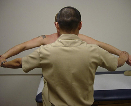
Empty-can test: evaluates the supraspinatus. The patient raises both arms slightly forward from the coronal plane of the trunk with thumbs pointing to the floor (as if emptying a can). The examiner applies pressure to the top of the arms, which the patient attempts to resist. Weakness indicates a supraspinatus tear. [Figure caption and citation for the preceding image starts]: Empty-can testFrom the collection of Daniel J. Solomon, MD; used with permission [Citation ends].
External rotation test: isolates the infraspinatus. With the arm at his or her side and the elbow flexed to 90°, the patient attempts to externally rotate against resistance supplied by the examiner. Infraspinatus tears result in pain and weakness. [Figure caption and citation for the preceding image starts]: External rotation testFrom the collection of Daniel J. Solomon, MD; used with permission [Citation ends].
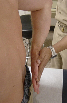
Lift-off test: evaluates the patient's ability to lift the hand away from the small of the back as the examiner applies resistance. The examiner must ensure that the patient uses the shoulder and arm rather than wrist and fingers to perform this task. Weakness suggests a subscapularis tear. [Figure caption and citation for the preceding image starts]: Lift-off testFrom the collection of Daniel J. Solomon, MD; used with permission [Citation ends].
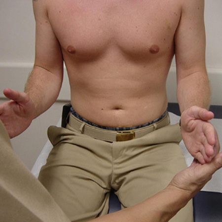
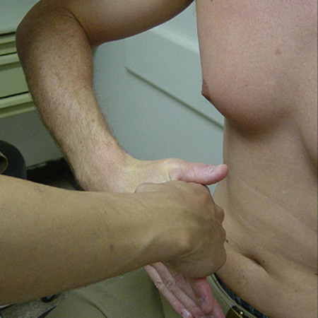
Belly-press test: the patient presses the hand against the umbilicus with the elbow forward from the trunk. The examiner applies resistance by placing his or her hand between the patient's hand and abdomen. Inability to maintain elbow anterior to the coronal plane of the trunk suggests a subscapularis tear. This test may also be performed supine with the examiner stabilising the scapula.[Figure caption and citation for the preceding image starts]: Belly-press testFrom the collection of Daniel J. Solomon, MD; used with permission [Citation ends].
The patient should be actively pushing against the examiner's hand in all these tests. Muscle strength can be graded on a 0 to 5 scale. Weakness (0/5 to 3/5) suggests a rotator cuff tear.
Provocative impingement tests (Neer and Hawkins)
Typically positive with rotator cuff tears. The Neer impingement test can be performed with the patient seated or standing. The examiner keeps one hand on the patient's scapula to prevent rotation. As the patient's arm is elevated by the examiner, reproduction of pain is a positive test for impingement. With the Hawkins test, the patient's arm is positioned at 90° of elevation and the elbow is bent to 90°. The examiner places an internal rotation force on the patient's arm. Reproduction of pain is a positive test for subacromial inflammation and rotator cuff tendinopathy.
Subacromial injection with local anaesthetic
In practice, the presence of pain and limited active motion, or signs of rotator cuff tendinopathy, warrants consideration of diagnostic subacromial injection with local anaesthetic followed by re-examination. If the rotator cuff is intact, and pain is the causal mechanism of apparent weakness, strength should improve after the pain-relieving injection.
Contribution of history and clinical tests to the diagnosis
Opinions differ on the importance of history and physical examination in diagnosing rotator cuff tear.[20]
One study of 103 patients with tears reported that the characteristics of the pain, the site of tenderness, and weakness to resisted abduction did not correlate with the presence or severity of the tear, although the extent of tear did relate to the limitation of shoulder abduction.[21] Similarly, one systematic review that evaluated two level IV studies (case control/cohort) found that historical findings (including reports of the presence or absence of pain at rest, pain during sleep, or pain during motion) did not help to identify patients with rotator cuff tear.[22] This systematic review also assessed the effect of physical examination manoeuvres and found that a positive external rotation resistance test was among the most accurate findings for detecting rotator cuff disease. Positive results on provocative tests (Neer test and Hawkins test) did not appear to be helpful.[22]
In another systematic review that evaluated the sensitivity and specificity of five clinical tests, the Hawkins-Kennedy test, the Neer sign, and the empty can test were better for ruling out subacromial irritation (when the examination was normal) than ruling it in.[23] The drop arm test and the lift-off test had higher pooled specificities than sensitivities, and were more useful at ruling in rotator cuff tendinopathy if the test was positive.[23]
Of note, clinical manoeuvres evaluated in published studies are typically performed by specialists on referred patients. It is uncertain whether examinations performed by generalists or primary care physicians would elicit the same results. As tests are refined and specificity for individual tendons is increased, the examination will likely have increasing influence on decision-making.[Figure caption and citation for the preceding image starts]: Neer impingement testFrom the collection of Daniel J. Solomon, MD; used with permission [Citation ends]. [Figure caption and citation for the preceding image starts]: Hawkins impingement testFrom the collection of Daniel J. Solomon, MD; used with permission [Citation ends].
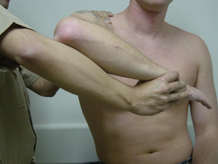 [Figure caption and citation for the preceding image starts]: Subacromial injection. Insert needle just inferior to posterior edge of acromion (x), aiming parallel to the undersurface of the acromionFrom the collection of Daniel J. Solomon, MD; used with permission [Citation ends].
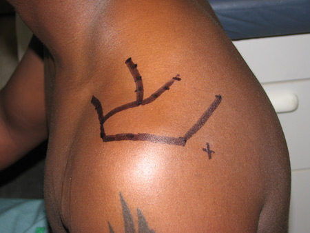
Imaging
X-rays are typically used during the initial evaluation to rule out fractures after trauma and to evaluate other pathology, such as acromioclavicular joint, glenohumeral arthritis, or rarely, a neoplasm.[17][18] Clinicians must be aware that apical lung tumours (Pancoast tumour) may present with shoulder pain.
Advanced imaging may be recommended if surgery is being contemplated or if a patient continues to have pain and decreased motion after at least 6 weeks of therapy.
Magnetic resonance imaging (MRI) and ultrasound are usually the advanced imaging modalities of choice to diagnose acute rotator cuff injury when x-rays are either negative or indeterminate, but the physical examination is consistent with rotator cuff tear.[17] These procedures are equivalent alternatives and only one needs to be ordered; however, MRI without contrast might be the preferred imaging modality if patients have a restricted range of motion due to acute pain, if there is suspicion of other intraarticular pathologies, such as labral tears, or for patients with large body habitus.[17]
One meta-analysis found 95% sensitivity and 96% specificity for the diagnostic accuracy of ultrasound for full-thickness rotator cuff tears, and 72% sensitivity and 93% specificity for partial-thickness tears.[24] However, the diagnostic accuracy of ultrasound is operator-dependent. Ultrasound is a better modality than MRI if there is prior metal hardware, such as suture anchors, present in the humeral head or glenoid.[17] It also allows a dynamic evaluation of the shoulder, whereas other imaging modalities are static.
Sensitivity and specificity of MRI utilised to detect full-thickness rotator cuff tears are 91% and 97%, respectively.[25] MRI and ultrasound provide the surgeon with important information to permit better preoperative planning and establish realistic treatment expectations. The amount of retraction and atrophy, tear size, number of tendons involved, and presence of fatty infiltration are all important considerations in determining a treatment strategy. In particular, the sagittal oblique images provide excellent information regarding muscle quality and infiltrate.
For patients with chronic shoulder pain due to suspected rotator cuff disorder based on the physical examination, advanced imaging usually includes ultrasound, MR arthrography, or MRI without contrast if the initial x-rays are normal or inconclusive.[18] These procedures are considered to be equivalent alternatives, but there is evidence to suggest that MR arthrography may be more effective for the diagnoses of both partial and full thickness tears.[26] In addition, ultrasound is technician dependent and may not detect incomplete tears.
Computed tomography (CT) scan and CT arthrography are less commonly used, as further pathology can be more difficult to identify with these modalities. In addition, they subject the patient to appreciable ionising radiation. However, they may be used if other imaging is not available. CT arthrograms have been shown to be comparable to MR arthrography in diagnosing full-thickness rotator cuff tears, but inferior to MR arthrography for diagnosing partial-thickness rotator cuff tears.[27]
Use of this content is subject to our disclaimer