Images and videos
Images
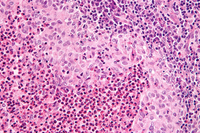


Langerhans cell histiocytosis
Very high magnification micrograph of Langerhans cell histiocytosis. H&E stain. It is characterized by Langerhans-type histiocytes that have a reniform (kidney-shaped) nucleus and stain with S100 and CD1a
Nephron. Reproduced under a creative commons license CC BY-SA 3.0: https://creativecommons.org/licenses/by-sa/3.0/deed.en
See this image in context in the following section/s:

Langerhans cell histiocytosis
Langerhans cell histiocytosis rash on an infant's abdomen
Reproduced with permission from Science Photo Library
See this image in context in the following section/s:
Langerhans cell histiocytosis
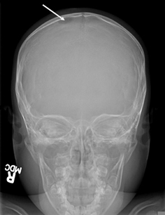
Skull x-ray showing lytic bone lesion in the right posterior parietal area of the skull
From the personal collection of Oussama Abla, MD; used with permission
See this image in context in the following section/s:
Use of this content is subject to our disclaimer