Aetiology
Aetiology
The causes of acute red eye can be considered within the following categories:[3]
Adnexal causes
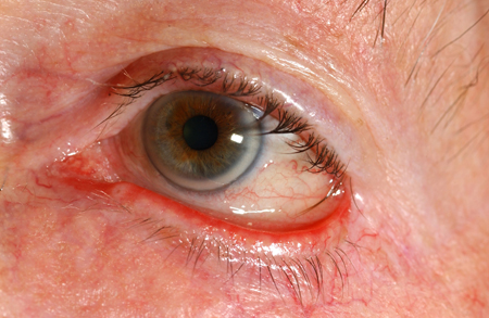
Trichiasis: posterior misdirection of the eyelashes from the normal site of origin [Figure caption and citation for the preceding image starts]: TrichiasisPrivate collection - courtesy of Mr Hugh Harris [Citation ends].

Entropion: inward turning of the eyelid margin [Figure caption and citation for the preceding image starts]: EntropionPrivate collection - courtesy of Mr Hugh Harris [Citation ends].

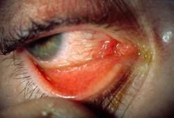
Ectropion: outward turning of the eyelid margin [Figure caption and citation for the preceding image starts]: EctropionPrivate collection - courtesy of Mr Hugh Harris [Citation ends].
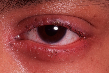
Blepharitis: inflammation of the eyelid margin [Figure caption and citation for the preceding image starts]: BlepharitisPrivate collection - courtesy of Mr Hugh Harris [Citation ends].
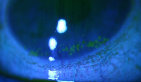
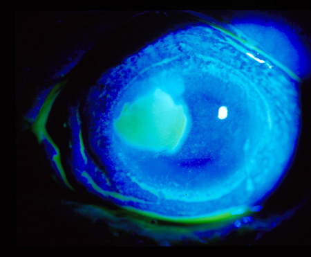
Dry eye: symptoms or signs consistent with a deficiency of the precorneal tear film. [Figure caption and citation for the preceding image starts]: Dry eye (stained with fluorescein)From the personal collection of Dr Jonathan Smith; used with permission [Citation ends].
Conjunctival causes
Bacterial conjunctivitis: inflammation of the conjunctiva caused by bacterial infection [Figure caption and citation for the preceding image starts]: Bacterial conjunctivitisPrivate collection - courtesy of Mr Hugh Harris [Citation ends].
Viral conjunctivitis: inflammation of the conjunctiva caused by viral infection. Some patients with COVID-19 may present with features typical of viral conjunctivitis.[4] Primary care physicians should maintain a high index of suspicion for this uncommon presentation[5][Figure caption and citation for the preceding image starts]: Viral conjunctivitisPrivate collection - courtesy of Mr Hugh Harris [Citation ends].
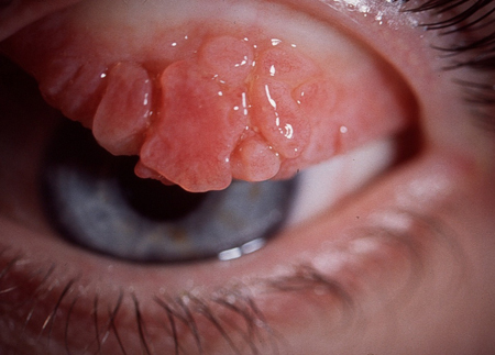
Allergic (vernal) conjunctivitis: inflammation of the conjunctiva occurring during an allergic response [Figure caption and citation for the preceding image starts]: Allergic (vernal) keratoconjunctivitisPrivate collection - courtesy of Mr Hugh Harris [Citation ends].
Neonatal conjunctivitis: inflammation of the conjunctiva within the first month of life
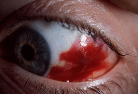
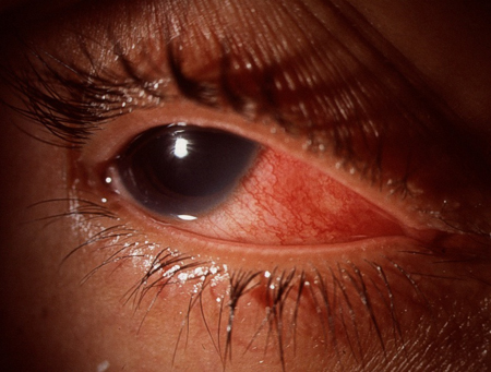
Subconjunctival haemorrhage [Figure caption and citation for the preceding image starts]: Subconjunctival haemorrhagePrivate collection - courtesy of Mr Hugh Harris [Citation ends].
Subtarsal foreign body [Figure caption and citation for the preceding image starts]: Subtarsal foreign body: vertical corneal abrasions seen with fluorescein stainPrivate collection - courtesy of Mr Hugh Harris [Citation ends].
Conjunctival foreign body.
Corneal causes
Bacterial corneal ulcer: corneal epithelial defect caused by bacterial infection [Figure caption and citation for the preceding image starts]: Corneal ulcer seen with fluorescein stainPrivate collection - courtesy of Mr Hugh Harris [Citation ends].
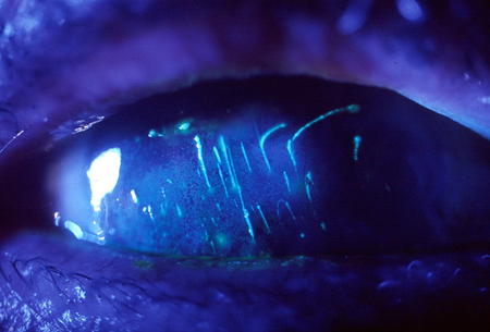
Viral corneal ulcer: corneal epithelial defect caused by viral infection [Figure caption and citation for the preceding image starts]: Dendritic ulcer seen with fluorescein stainPrivate collection - courtesy of Mr Hugh Harris [Citation ends].

Fungal corneal ulcer: corneal epithelial defect caused by fungal infection
Contact lens-related
Corneal foreign body [Figure caption and citation for the preceding image starts]: Corneal foreign bodyPrivate collection - courtesy of Mr Hugh Harris [Citation ends].
Corneal abrasion: corneal epithelial defect usually caused by trauma. [Figure caption and citation for the preceding image starts]: Corneal abrasion seen with fluorescein stainPrivate collection - courtesy of Mr Hugh Harris [Citation ends].

Inflammatory causes
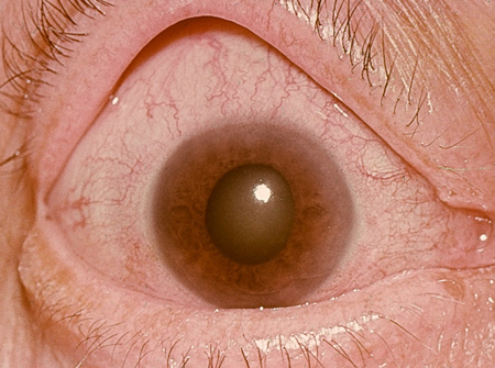
Anterior uveitis: inflammation of the anterior portion of the uveal tract [Figure caption and citation for the preceding image starts]: Anterior uveitis with posterior synechiaePrivate collection - courtesy of Mr Hugh Harris [Citation ends].

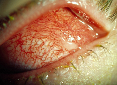
Scleritis: inflammation of the sclera [Figure caption and citation for the preceding image starts]: ScleritisPrivate collection - courtesy of Mr Hugh Harris [Citation ends].
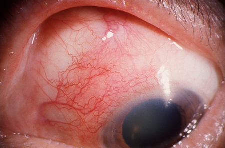
Episcleritis: inflammation of the episclera. [Figure caption and citation for the preceding image starts]: EpiscleritisPrivate collection - courtesy of Mr Hugh Harris [Citation ends].
Traumatic causes
Physical [Figure caption and citation for the preceding image starts]: Penetrating corneal injury with iris prolapsePrivate collection - courtesy of Mr Hugh Harris [Citation ends].
Chemical.
Other
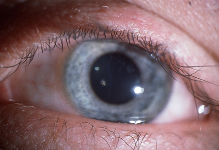
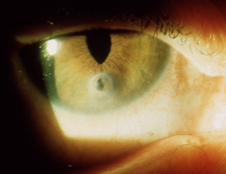
Angle-closure glaucoma: closure of the iridocorneal angle leading to an acute rise in intra-ocular pressure. [Figure caption and citation for the preceding image starts]: Angle-closure glaucoma: central corneal oedema with an oval-shaped mid-dilated pupil.Private collection - courtesy of Mr Hugh Harris [Citation ends].
Most common conditions
Those commonly presenting to a primary care physician are:
Infective conjunctivitis[6][Figure caption and citation for the preceding image starts]: Bacterial conjunctivitisPrivate collection - courtesy of Mr Hugh Harris [Citation ends].
Allergic conjunctivitis [Figure caption and citation for the preceding image starts]: Allergic (vernal) keratoconjunctivitisPrivate collection - courtesy of Mr Hugh Harris [Citation ends].
Dry eye and other adnexal problems.[7][8][Figure caption and citation for the preceding image starts]: TrichiasisPrivate collection - courtesy of Mr Hugh Harris [Citation ends].
[Figure caption and citation for the preceding image starts]: EntropionPrivate collection - courtesy of Mr Hugh Harris [Citation ends].[Figure caption and citation for the preceding image starts]: EctropionPrivate collection - courtesy of Mr Hugh Harris [Citation ends].[Figure caption and citation for the preceding image starts]: BlepharitisPrivate collection - courtesy of Mr Hugh Harris [Citation ends].[Figure caption and citation for the preceding image starts]: Dry eye (stained with fluorescein)From the personal collection of Dr Jonathan Smith; used with permission [Citation ends].
Sight-threatening causes
Causes of red eye that can threaten vision by leading to reduced visual acuity include:
Angle-closure glaucoma [Figure caption and citation for the preceding image starts]: Angle-closure glaucoma: central corneal oedema with an oval-shaped mid-dilated pupil.Private collection - courtesy of Mr Hugh Harris [Citation ends].
Chemical injuries
Conditions affecting the cornea
Trauma
Anterior uveitis. [Figure caption and citation for the preceding image starts]: Anterior uveitis with posterior synechiaePrivate collection - courtesy of Mr Hugh Harris [Citation ends].
Causes of red eye that can threaten vision by leading to globe rupture or perforation include: [Figure caption and citation for the preceding image starts]: Penetrating corneal injury with iris prolapsePrivate collection - courtesy of Mr Hugh Harris [Citation ends].
Scleritis [Figure caption and citation for the preceding image starts]: ScleritisPrivate collection - courtesy of Mr Hugh Harris [Citation ends].
Physical trauma
Corneal ulceration
High-velocity foreign bodies.
These are discussed further with initial management in Emergencies: Urgent Considerations.
Risk factors
Risk factors associated with specific causes of red eye include:
Anterior uveitis: [Figure caption and citation for the preceding image starts]: Anterior uveitis with posterior synechiaePrivate collection - courtesy of Mr Hugh Harris [Citation ends].
human leukocyte antigen-B27 histocompatibility complex-positive patients, tuberculosis, syphilis, Lyme disease, sarcoidosis, Behcet's disease, and pauciarticular juvenile chronic arthritis.Scleritis: [Figure caption and citation for the preceding image starts]: ScleritisPrivate collection - courtesy of Mr Hugh Harris [Citation ends].
connective tissue disorders including rheumatoid arthritis, granulomatosis with polyangiitis (formerly known as Wegener's granulomatosis), systemic lupus erythematosus (SLE), and relapsing polychondritis.Episcleritis: [Figure caption and citation for the preceding image starts]: EpiscleritisPrivate collection - courtesy of Mr Hugh Harris [Citation ends].
connective tissue disorders including rheumatoid arthritis, granulomatosis with polyangiitis (formerly known as Wegener's granulomatosis), and SLE.Angle-closure glaucoma: [Figure caption and citation for the preceding image starts]: Angle-closure glaucoma: central corneal oedema with an oval-shaped mid-dilated pupil.Private collection - courtesy of Mr Hugh Harris [Citation ends].
hypermetropia, drugs (e.g., therapeutic mydriatics, drugs with unwanted mydriatic effects such as systemic anticholinergics and topiramate).Subconjunctival haemorrhage: [Figure caption and citation for the preceding image starts]: Subconjunctival haemorrhagePrivate collection - courtesy of Mr Hugh Harris [Citation ends].
hypertension, systemic anticoagulation, bleeding abnormalities (leukaemia, clotting disorders), conjunctival vascular lesion, trauma (including contact lens-related injury), and diabetes.Dry eye: [Figure caption and citation for the preceding image starts]: Dry eye (stained with fluorescein)From the personal collection of Dr Jonathan Smith; used with permission [Citation ends].
connective tissue disorders including Sjogren's syndrome, rheumatoid arthritis, and SLE.
Use of this content is subject to our disclaimer