Investigations
1st investigations to order
cranial CT
Test
Use the criteria listed in the two flowcharts below to carefully identify who to send for a computed tomography (CT) scan and the appropriate timeframe required to exclude serious complications.[28]
Although skull fractures often present with no clinical symptoms or signs on physical examination, they are significant risk factors for intracranial pathology.
Any patient who is considered high-risk for brain injury and/or cervical spine injury or has deteriorating neurological status must have a CT scan within 1 hour of the risk being identified.[28]
If the patient has sustained a head injury and has no other indications for a CT head scan, but is on anticoagulant treatment or antiplatelet treatment (including vitamin K antagonists, direct-acting oral anticoagulants [DOACs], heparin and low molecular weight heparins) or antiplatelet treatment (excluding aspirin monotherapy), consider:[28]
A CT scan within 8 hours of the injury (for example, if it is difficult to carry out a risk assessment or if the patient might not return to the emergency department if they have signs of deterioration)
A CT scan within the hour if the patient presents more than 8 hours after the injury.
Patients who have no other risk factors for brain injury but are taking anticoagulants or antiplatelet treatment (excluding aspirin monotherapy) have an increased risk of bleeding after a head injury.[28]
[Figure caption and citation for the preceding image starts]: Selection of adults for CT head scan; GCS = Glasgow Coma Scale; CT = computed tomography; A&E = accident and emergency departmentAdapted from Rajesh S et al. BMJ 2023;381:p1130 [Citation ends].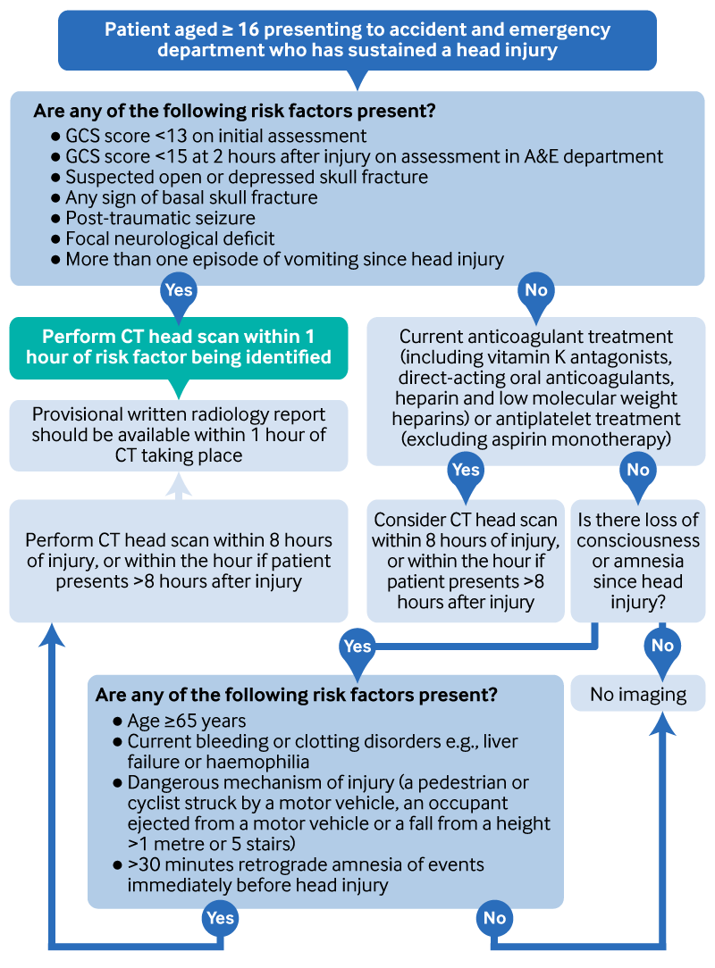 [Figure caption and citation for the preceding image starts]: Selection of children for CT head scan; GCS = Glasgow Coma Scale; CT = computed tomography; A&E = accident and emergency departmentAdapted from Rajesh S et al. BMJ 2023;381:p1130 [Citation ends].
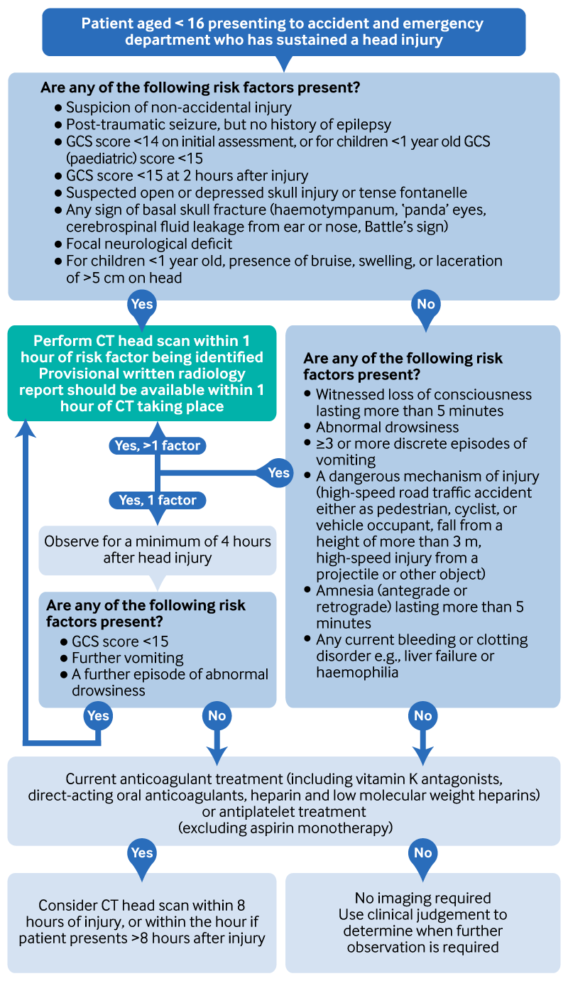
CT is superior to MRI for detecting skull fracture in both children and adults.[35][36][37]
Basilar skull fractures are the most difficult to detect; CT scans should be performed with thin cuts and should include three-dimensional reconstruction of some type.[38][39][40][41]
A retrospective comparison of three different reconstructive techniques revealed the best sensitivity with high-resolution multiplanar reformation (HRMPR), which is currently the standard of care, in combination with maximum intensity projection (MIP) reconstructions.[40] MIP reconstructions increase detection rate by 18% and can detect different types of fractures compared with HRMPR.[41]
Fracture detection is improved if more than one radiologist reviews the images.[40]
Prospective evaluation of several head trauma imaging guidelines found that protocols that increased sensitivity for detecting pathology were also associated with a significant number of unnecessary CT scans.[7]
The predictive value of many of the imaging criteria detailed above from the National Institute for Health and Care Excellence (NICE) in the UK were confirmed in a meta-analysis of 71 studies. This found that seizure, persistent vomiting, and coagulopathy all significantly predicted positive head CT findings in patients with mild brain injury.[42]
If physical abuse is suspected in a child, recommendations from the Royal College of Radiology in the UK are as follows:[43]
Imaging should always include skeletal survey in children aged under 2 years and skeletal survey and CT head scan in children under 1 year.
Children who are older than 1 year and have external evidence of head trauma and/or abnormal neurological symptoms or signs should also have a CT head scan.
Evidence: Decision rules for performing a CT scan in adults
There are multiple clinical decision rules to identify which adults should have a CT scan to determine whether they have clinically significant brain injury. The results of validation studies vary and there is no clear decision rule that should be used internationally. In the UK, follow the recommendations from the National Institute for Health and Care Excellence (NICE).
The NICE clinical decision rule for CT scanning adults with head injury combines patient selection for imaging with urgency and is based on the high- and medium-risk criteria of the Canadian CT Head Rule (CCHR).[28]
An update (2023) to the 2014 NICE guideline found no new evidence of sufficiently high quality to change the previous recommendation on clinical decision for CT scanning.[28]
NICE identified 33 diagnostic accuracy studies in adults, but no diagnostic randomised controlled trials. Most of the trials were in adults with mild traumatic brain injury, and the evidence for the majority of outcomes were assessed by GRADE as low to very low quality.
The updated evidence confirmed previous findings that the CCHR has good sensitivity (≥90%) when used as intended but in general has poor specificity (<60%).
The National Emergency X-Radiography Utilisation Study II (NEXUS II) decision rule performed similarly to CCHR; however, the evidence was more limited. Other decision rules had a similar sensitivity but lower specificity.
The CCHR was also the most cost effective of the decision rules assessed, further supporting its use as the basis for the NICE recommendations. There was no cost effectiveness evidence directly assessing the NICE 2014 rule.
A separate recommendation for people on anticoagulant or antithrombotic therapy, introduced in 2014, also remained unchanged.
In the 2023 NICE guideline update, only one study was identified comparing the performance of different decision rules in the same population of adults. This was a prospective diagnostic accuracy study from 2018, and it compared four decision rules (including the NICE 2014 decision tool).[28]
This study included 4557 adults with mild traumatic brain injury (six centres in the Netherlands). It found the NICE decision tool had a higher specificity but lower sensitivity compared with the CT in head injury patients (CHIP) rule, New Orleans Criteria (NOC), or CCHR.
Sensitivity for any intracranial injury on CT ranged from 73% with NICE to 99% with NOC; specificity ranged from 4% with NOC to 61% with NICE.
Sensitivity for potential neurosurgical lesions ranged between 85% with NICE and 100% with NOC; specificity ranged from 4% with NOC to 59% with NICE.
Of note, the sensitivity of the CCHR in this study was considerably lower than in other studies (<90%).
As the NICE 2014 decision tool was largely based on the CCHR, the NICE guideline committee postulated that there may be some differences in this study population, affecting the sensitivity of both rules; and that the lower sensitivities of the NICE tool in this study did not match their clinical experience
Evidence: Decision rules for performing a CT scan in children
Use the UK National Institute for Health and Care Excellence (NICE) criteria to identify which children and infants should have a CT scan to determine whether they have clinically significant brain injury. However, there is a paucity of externally validated studies and therefore NICE acknowledges that its recommendations should be used alongside clinical judgement.
The NICE clinical decision rule for CT scanning children and infants with head injury is based on the Children’s Head Injury Algorithm for the Prediction of Important Clinical Events (CHALICE).[28]
An update (2023) to the 2014 NICE guideline found no new evidence of sufficiently high quality to change the previous recommendation on clinical decision for CT scanning in children and infants.
NICE identified 42 diagnostic accuracy studies in children and infants, but no diagnostic randomised controlled trials. Most of the trials were in children and infants with mild traumatic brain injury, and the evidence for the majority of outcomes were assessed by GRADE as low to very low quality.
The updated evidence (n >40,000) for the CHALICE rule showed that it had >90% sensitivity and >80% specificity for clinically important injuries or neurosurgical outcomes.
There was some evidence that the Pediatric Emergency Care Applied Research Network (PECARN) and the Canadian Assessment of Tomography for Childhood Head Injury 7-item (CATCH‑7) rules may have slightly better sensitivity compared with CHALICE, especially for any severity of head injury. However, as the specificity of CHALICE was better than for other rules, the guideline panel did not feel any changes were required to the NICE 2014 criteria.
The committee also felt that the inclusion of timings in the NICE 2014 criteria, and their applicability to a more general population, made the NICE 2014 criteria more useful clinically than PECARN or CATCH-7.
Three studies assessed the National Emergency X-Radiography Utilisation Study II (NEXUS II) in children.[44][45][46] Sensitivity was >98% for any severity of injury, clinically important injuries, and neurosurgery outcomes. However, specificity was <50% for all outcomes, and only the outcome of clinically important injuries was assessed in a sufficiently large population.[28]
One small study was identified which assessed the Pittsburgh Infant Brain Inventory Score (n=891; infants aged 30 days to 1 year).[47] Using a cut-off score of ≥2, sensitivity was 93% for any severity of injury, although specificity was only 53%.[28] The rule had not been externally validated at the time of publication of the NICE 2023 guideline.
Evidence: CT head scan in patients on anticoagulant treatment with no other indications for CT
Observational evidence supports the recommendations made by the UK National Institute for Health and Care Excellence (NICE) to consider a CT scan in patients with head injury and no other risk factors for clinically significant brain injury but who are on anticoagulants or antiplatelets (other than aspirin monotherapy).
In the 2014 NICE head injury guideline, the guideline committee found no clinical decision rules for patients who have no history of amnesia or loss of consciousness who are on anticoagulant or antiplatelet therapy. As part of the 2023 update, the guideline committee looked for prognostic evidence from cohort studies in people on anticoagulant treatment.[28]
Adults
NICE identified five cohort studies in adults on anticoagulants only, and five in adults on anticoagulants and antiplatelet therapy.[28]
There was conflicting evidence as to whether people on anticoagulant or antiplatelet therapy are at an increased risk of intracranial haemorrhage. The guideline committee decided that the recommendation should be changed from 'request a CT scan' to 'consider a CT scan' to allow for shared decision making and for selected patients to be discharged without a CT scan.[28]
When considering the risks of not requesting a scan in all patients with mild head injury on anticoagulant or antiplatelet therapy without any other indication, the committee noted that delayed recovery was more likely than death if an intracranial haemorrhage was missed at initial presentation.
Further, for patients aged >74 years the committee felt that the risks of neurosurgical intervention may outweigh the benefits, and that this should also be taken into consideration when deciding whether to scan patients with no other risk factors.
The 2014 NICE guideline strategy of scanning all people on anticoagulants was not found to be cost effective in the base case analysis. However, sensitivity analyses found small changes in rates of admission or in assumptions about the effectiveness of immediate versus delayed neurosurgery meant it would be cost effective.[48][28]
In 2014, NICE made a research recommendation regarding people on antiplatelet therapy. At the 2023 update the decision was made, based on limited new evidence and clinical experience, to include patients on antiplatelet therapy in the same recommendation.[28]
The evidence for people on aspirin monotherapy was limited. Based on expert knowledge and clinical experience, the committee felt that the risk of intracranial haemorrhage in this population was low, therefore this population was excluded from the recommendation.
The guideline committee also discussed timing of imaging.
The evidence review in 2014 found that the median time in the study from injury to CT scan was 234 minutes (interquartile range 175 to 335 minutes) for patients diagnosed with an intracranial lesion at the first scan.
Therefore, a timeframe of within 8 hours of injury (giving time to detect a possible slow bleed) was recommended.
At the 2023 update, the committee agreed that the 2014 recommendations could also be applicable to people presenting >8 hours post-injury based on their clinical experience and from extrapolating the data from <8 hours. The committee felt that in this situation the CT scan should be done within the hour.[28]
Children and infants
For children and infants, the 2014 guideline group identified one prospective cohort study (43,904 children under 18 years) with non-trivial blunt head trauma.[49]
Only 15 children were taking anticoagulation therapy and only two children in the entire study population were diagnosed with an intracranial haemorrhage, of which one was taking warfarin.
No new evidence for children and infants was identified at the 2023 update.
Due to the very limited evidence in children, the decision was made to extend the recommendation based on the evidence in adults to anyone on anticoagulants or antiplatelets.
Due to the lack of evidence on risk of intracranial haemorrhage in people with a pre-injury coagulopathy (including patients on anticoagulant or antiplatelet therapy) and no other risk factors, the guideline committee made a research recommendation in this area.[28]
Adjuncts to conventional CT include the use of:
Intrathecal contrast to localise the source of cerebrospinal fluid leak[37]
CT angiography (CTA) if there is any suspicion for vascular injury, such as when the fracture involves the carotid canal or overlies a vessel (e.g., middle meningeal artery, sagittal sinus).[37] Use the expanded Denver criteria to determine if CTA is necessary.[50][51]
Practical tip
A radiologist (ST3 or higher in the UK) should be present at the trauma call to verbally report the CT scan immediately (with a written report within 1 hour). If the radiologist identifies a fracture involving a vascular foramen, the patient should proceed to have a CT angiogram while still in the scanner.
Result
detects skull fractures and any associated intracranial pathology
Investigations to consider
skeletal survey
Test
Conduct a skeletal survey if child abuse is a suspected underlying aetiology.[28][43]
Suspect child maltreatment if a child has one or more fractures in the absence of a medical condition that predisposes to fragile bones (for example, osteogenesis imperfecta, osteopenia of prematurity) or if the explanation is absent or unsuitable. Presentations include:[14]
Fractures of different ages
X-ray evidence of occult fractures (fractures identified on x-rays that were not clinically evident), e.g., rib fractures in infants.
The skeletal survey should be acquired and reported within 24 hours and certainly no later than 72 hours from the request being made.[43]
Imaging of hands, feet, long bones, skull, spine, and ribs (including oblique ribs) should be performed, with high-definition imaging (CT/MRI of evident fractures) if possible. Plain x-rays of the skull are a useful part of the skeletal survey in children presenting with suspected non-accidental injury.[28]
Although skeletal survey is specifically recommended in children aged under 2 years where child abuse is suspected, skeletal survey may also occasionally be indicated in older children; this should be considered on a case-by-case basis. This may include children with communication or learning difficulties or neurodisability who may be unable to give a history of physical abuse or children where there is a clinical suspicion of skeletal injury.[43]
All children should have follow-up imaging.[43] The repeat skeletal survey should be performed ideally within 11 to 14 days, and no later than 28 days, after the initial investigation.[43] The repeat survey will give further information about ambiguous findings, identify further fractures, and add information about the age of a fracture.[43]
Ensure a clinician with expertise in non-accidental injuries in children is involved in any suspected case of non-accidental injury in a child.[28]
Result
variable; unexplained fractures including skull fractures, long-bone fractures, rib fractures, and classic metaphyseal lesions
MRI
Test
Do not routinely perform an MRI for the evaluation of skull fractures.
If you suspect non-accidental injury in a child, perform an MRI.
MRI can be a useful adjunct or a secondary imaging modality, particularly if there is continuing concern of intracranial pathology in the absence of CT findings.[28]
Its main benefit is increased detection of associated intracranial pathology. MRI can detect diffuse axonal injury not seen on the CT scan, and can increase detection of intracranial haemorrhage (extradural/subdural) by up to 30%.[4][35][39][52]
MRI (alone or in conjunction with MR angiography) may also be useful if the fracture involves major vascular structures (e.g., the carotid canal or superior sagittal sinus), to assess underlying vascular injury/pathology.[37][39][53][54][55]
Result
detects skull fractures and any associated intracranial pathology
MR angiography
Test
MR angiography (MRA) may be useful if the fracture involves major vascular structures (e.g., the carotid canal or superior sagittal sinus), to assess underlying vascular injury/pathology.[37][39][53][54][55] Use the expanded Denver criteria to determine if MRA is necessary.[50][51]
Result
detects any associated vascular injury/pathology
beta-2 transferrin assay
Test
For any patient with head trauma and otorrhoea/rhinorrhoea (if clear or blood-tinged drainage is present from the nose or ears), request a beta-2 transferrin assay of the suspect fluid.
If positive, it indicates cerebrospinal fluid (CSF) leakage and is reliable even in the presence of blood or mucus. It has a sensitivity of nearly 100% and a specificity of 95%.[56]
Result
positive if CSF leak
plain skull x-ray
Test
Do not routinely request an x-ray before discussing with a neuroscience unit.[28]
Plain films were previously used to help screen patients who would benefit from CT scanning. However, they offer no additional information and are associated with poor sensitivity and failure to detect any associated intracranial pathology.[36] With the widespread availability of CT scans to help detect intracranial pathology, plain skull x-rays are no longer recommended as a first-line investigation.
Plain x-rays of the skull are a useful part of the skeletal survey in children presenting with suspected non-accidental injury.[28]
Skull x-rays may be used as an interim aid if CT scanning is not available
Result
may reveal fracture
clotting screen
Test
Consider a clotting screen (prothrombin time, partial thromboplastin time, and INR) in patients taking anticoagulants.
Result
may be normal; may show coagulopathy
Use of this content is subject to our disclaimer