Approach
The most useful tool in the diagnosis of carpal tunnel syndrome (CTS) is a combination of characteristic clinical findings (particularly symptoms) plus abnormalities on electromyography (EMG) testing.
History
A history of gradual onset, intermittent (bilateral) hand numbness or pain, with waking up at night-time, relieved by shaking or flicking the wrist, is more than 90% specific for CTS.[26] Numbness is typically confined to the palmar aspect of the thumb and radial fingers (excluding the little finger). On awakening, patients may have difficulty extending or flexing fingers.
In the daytime symptoms are usually associated with activity, notably holding a book or newspaper or telephone, driving, or using a wrench or screwdriver. The patient may also complain of clumsiness, especially during fine-motor tasks and weakness. Some may have associated vasomotor symptoms and complain of cold sensitivity.
Involvement of both hands is helpful in making the diagnosis, as several of the potential differential diagnoses (radiculopathy, plexopathy, stroke, and proximal median neuropathies) are nearly always unilateral.
Patients with sensory symptoms just in the little finger (likely to be ulnar neuropathy), sensory symptoms proximal to the wrist crease (likely to be radiculopathy or more proximal neuropathies), or just pure hand weakness and wasting (possible motor neuron disease) are unlikely to have CTS. Typically, patients often complain of 'numbness' in the forearm and arm with CTS; however, on closer questioning it usually becomes apparent that patients are describing an aching or painful limb rather than proximal sensory nerve symptoms.
Musculoskeletal examination
The musculoskeletal examination of the cervical spine and upper extremity is often more useful than the neurological examination, given that many musculoskeletal disorders co-exist or are confused with CTS. Weakness of the thenar muscles is a common finding. However, atrophy of the thenar eminence is a sign of severe CTS.
Since radiculopathy is a common differential diagnosis, examination of the neck (range of motion and Spurling's maneuver) can help to confirm or rule out radiculopathy.
Patients should be examined for De Quervain's tenosynovitis, osteoarthritis of the first carpometacarpal joint, and flexor and extensor tendonitis at the wrist. These conditions may be the cause of the patient's main complaint or may be contributing to compression of the median nerve. Focal swelling near the carpal tunnel and/or complaints of pain in the non-dominant hand may indicate a space-occupying lesion. [Figure caption and citation for the preceding image starts]: Weak abduction of the thumb (4/5) on both sides but no thenar wastingFrom the personal collection of Dr Nigel Ashworth; used with permission [Citation ends].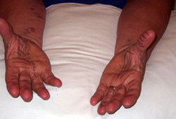
Several specific tests for CTS have been described, including Tinel's sign and Phalen's test. Tinel's sign has been shown to be present in up to 40% of people without CTS, and the sensitivity of Phalen's test ranges from 10% to 90%.These tests are not recommended as useful.[27]
Neurological examination
The neurological examination is often most useful in ruling out other conditions (such as motor neuron disease or radiculopathy). Motor strength and bulk of the thenar muscles are unreliable signs, and reflex examination is only useful in ruling out other diagnoses (i.e., typically normal in CTS).
Sensory abnormalities to pinprick that are predominantly over the palmar aspect of the first 3 fingers and lateral half of the fourth, with sparing of the thenar eminence, is the most useful finding (median nerve distribution). However, typically these areas are also the most heavily calloused.[Figure caption and citation for the preceding image starts]: Typical area of diminished pinprick sensation (posterior) in CTSFrom the personal collection of Dr Nigel Ashworth; used with permission [Citation ends]. [Figure caption and citation for the preceding image starts]: Typical area of diminished pinprick sensation (anterior) in CTS; note sparing of thenar eminence (supplied by palmar branch of median nerve), and that right hand has more extensive loss than leftFrom the personal collection of Dr Nigel Ashworth; used with permission [Citation ends].
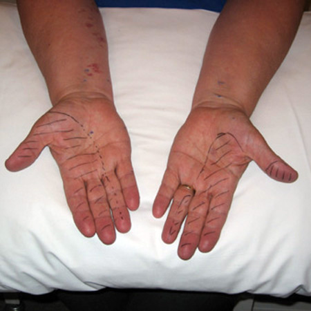
Diagnostic tests
The most sensitive and specific test is EMG (variously called electromyography, neurophysiology, electrodiagnostic testing, peripheral nerve conduction tests, electrophysiology, and clinical neurophysiology), which is usually widely available. It is recommended in all patients with a clinical diagnosis of CTS, as it is easy to perform, well tolerated, can confirm and localise damage to the median nerve in the carpal tunnel, and can categorise severity of damage to the nerve that helps to guide management.[28][29][30][31] Additionally, the EMG study can rule out other diagnoses (e.g., C6 radiculopathy) or provide evidence of another co-existent disease (e.g., polyneuropathies). It provides an objective baseline that can be followed over time to indicate success of treatment or progression. Patients with normal studies do worse with surgical release, and those in the severe category do poorly with conservative management. Some clinicians offer conservative initial treatment with splints for patients with typical symptoms and signs of CTS, followed by EMG if the symptoms fail to resolve.
Many electrodiagnostic laboratories are now using ultrasonography of the median nerve as a complementary test for CTS. Ultrasound can identify any structural abnormalities that might be impacting the nerve (such as a ganglion cyst or tendinitis) and can also assist in the diagnosis of CTS given that the median nerve might swell and become enlarged when it is damaged.[32] In addition, ultrasound can assist in guiding needle placement for steroid injection into the carpal tunnel.[33][34]
High-resolution ultrasound or magnetic resonance imaging (depending on local availability) of the wrist is useful if a space-occupying lesion is suspected.[32] Abnormalities in the shape and dimensions of the median nerve have been described at a variety of locations along the carpal tunnel (some of these abnormalities have also been described in people without CTS).
Use of this content is subject to our disclaimer