History and exam
Key diagnostic factors
common
history of traumatic or nontraumatic cutaneous lesion
anesthesia or severe pain over site of cellulitis
fever
palpitations, tachycardia, tachypnea, hypotension, and lightheadedness
uncommon
vesicles or bullae
Examination of the skin overlying the area of cellulitis may reveal vesicles or bullae.[3][16] It should be noted that patients with necrotizing fasciitis can present with normal overlying skin and that skin changes overlying group A streptococcal necrotizing fasciitis are a late sign.[16] Subtle skin changes such as leakage of fluid and edema precede the overt skin changes of blistering and redness. [Figure caption and citation for the preceding image starts]: Small areas of skin necrosis in a young woman with cellulitis and necrotizing fasciitis of her lower abdomen 5 days after a cesarean sectionFrom: Hasham S, Matteucci P, Stanley PRW, et al. Necrotising fasciitis. BMJ. 2005 Apr 9;330(7495):830-3 [Citation ends].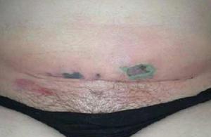
gray discoloration of skin
Examination of the skin overlying the area of cellulitis may reveal grayish discoloration. It should be noted that patients with necrotizing fasciitis can present with normal overlying skin and that skin changes overlying group A streptococcal necrotizing fasciitis are a late sign.
edema or induration
Examination of the skin overlying the area of cellulitis may reveal edema.[5] Induration may be noted beyond the area of cellulitis.[5] It should be noted that patients with necrotizing fasciitis can present with normal overlying skin and that skin changes overlying group A streptococcal necrotizing fasciitis are a late sign. Subtle skin changes such as leakage of fluid and edema precede the overt skin changes of blistering and redness. [Figure caption and citation for the preceding image starts]: Small areas of skin necrosis in a young woman with cellulitis and necrotizing fasciitis of her lower abdomen 5 days after a cesarean sectionFrom: Hasham S, Matteucci P, Stanley PRW, et al. Necrotising fasciitis. BMJ. 2005 Apr 9;330(7495):830-3 [Citation ends].[Figure caption and citation for the preceding image starts]: Necrotizing fasciitis on the right abdomen of a 2-year old girl following varicella infectionFrom: de Benedictis FM, Osimani P. Necrotising fasciitis complicating varicella. BMJ Case Rep. 2009;2009:bcr2008141994 [Citation ends].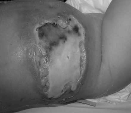
location of lesion
About half of cases occur in the extremities, with the remainder concentrated in the perineum, trunk, and head and neck areas.[1][5][16][19][20] The most common site of group A streptococcal necrotizing fasciitis is the thigh and necrotizing fasciitis of a limb, especially the arm, is more likely to be due to group A streptococcus than a polymicrobial infection. Some cases of necrotizing fasciitis may have associated myositis due to contiguous spread. This is more common in group A streptococcal than polymicrobial infections.
Necrotizing fasciitis in the context of recent abdominal surgery or in the groin is most likely to be polymicrobial.
Use of this content is subject to our disclaimer