Tests
1st tests to order
CXR
Test
Chest radiography is the first-line imaging modality in any patient presenting with known trauma.[33][34] This not only helps detect the actual rib fracture, but pneumothorax, hemothorax, and aortic injury can also be rapidly evaluated. However, conventional chest radiographs can miss up to 50% of rib fractures.[35] In practice, a single rib fracture might be diagnosed clinically based on symptoms and a clear history of thoracic trauma, even if no fracture is identified on CXR.
It is not usually necessary to perform dedicated rib radiography, in addition to chest radiography, for the diagnosis of rib fractures in adults after minor trauma.[33]
Pneumothorax occurs in about 14% to 37% of rib fractures, hemopneumothorax in 20% to 27%, pulmonary contusions in 17%, and a flail chest in up to 6%.[8][9][10][Figure caption and citation for the preceding image starts]: CT scan showing large left-sided pneumothoraxFrom the collection of Dr Paul Novakovich; used with permission [Citation ends].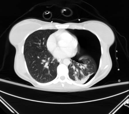 [Figure caption and citation for the preceding image starts]: CXR depicting the same pneumothorax as shown on CTFrom the collection of Dr Paul Novakovich; used with permission [Citation ends].
 [Figure caption and citation for the preceding image starts]: CXR showing multiple posterior left-sided rib fracturesFrom the collection of Dr Paul Novakovich; used with permission [Citation ends].
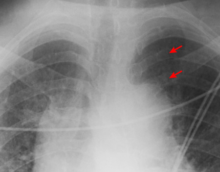
Result
rib fracture(s) ± associated parenchymal injury; pneumothorax; effusion/hemothorax
x-ray pelvis
Test
Typically, patients presenting after high-energy trauma should receive an immediate chest and pelvic radiograph to rule out life-threatening injury.[39]
Result
may reveal other injuries
Tests to consider
CT chest
Test
Conventional chest radiographs can miss up to 50% of rib fractures.[35] CT of the chest has much greater sensitivity in detecting rib fractures as well as other injuries.[41]
Should be considered if the patient sustained major trauma or if a CXR is negative for rib fracture, clinical features are suggestive of fracture, and confirmation of a fracture would change the patient’s management.
In minor trauma, the increased sensitivity of CT does not necessarily alter management or clinical outcomes of patients who do not have associated injuries.[33][Figure caption and citation for the preceding image starts]: CT scan showing bilateral posterior rib fracturesFrom the collection of Dr Paul Novakovich; used with permission [Citation ends].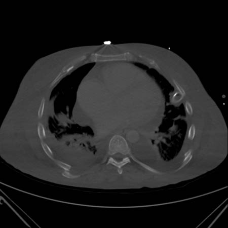 [Figure caption and citation for the preceding image starts]: CT showing right anterolateral rib fractureFrom the collection of Dr Paul Novakovich; used with permission [Citation ends].
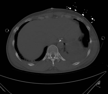 [Figure caption and citation for the preceding image starts]: CT showing left posterior segmental rib fractureFrom the collection of Dr Paul Novakovich; used with permission [Citation ends].
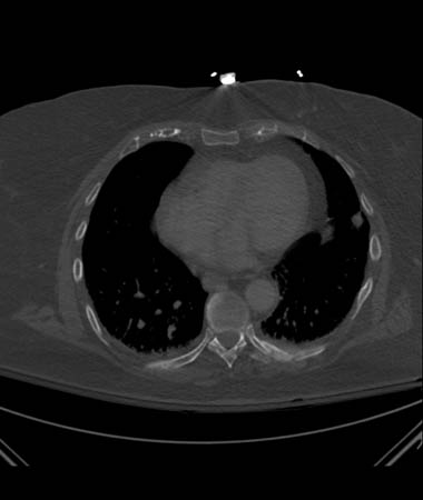
Result
rib fracture(s) ± associated parenchymal injury; sternal fracture; pneumothorax; hemothorax; great vessel injury; pneumomediastinum; thoracic vertebral fractures
ultrasound chest
angiography
Test
Traumatic injuries to the first rib have been associated with concomitant great-vessel injury in 3% of patients.[23]
Routine use of angiography is not indicated in the absence of discrepant pulses, mediastinal widening on chest x-ray, brachial plexus injury, or an expanding hematoma.[23]
Result
may reveal injury to any of the great vessels
CT of head, cervical spine, chest, abdomen, and pelvis
Test
Typically, patients presenting after high-energy trauma should receive an immediate chest and pelvic radiograph to rule out life-threatening injury.[39] If the patient sustained major trauma and is stable, a CT scan of the head, cervical spine, chest, abdomen, and pelvis may be performed in adults to rule out other injury.[39]
Result
may reveal other injuries
skeletal survey (children)
Test
Any rib fracture in a child or infant should be assumed to be the result of physical abuse until shown otherwise.
A skeletal survey should be considered in all children with suspected physical abuse.
Oblique views of the ribs are recommended in cases where abuse is suspected.[40] Oblique views increase diagnostic accuracy for rib fractures, which are strong positive predictors of abuse and may be the only skeletal manifestation.
A referral to child protection services should be considered.
Result
may reveal other injuries
Use of this content is subject to our disclaimer