Early diagnosis is essential for prompt therapeutic decisions and prevention of complications. Diagnosis is based on the signs and symptoms of inflammation in the presence of peritonitis localized to the right upper quadrant (RUQ) of the abdomen.[7]Indar AA, Beckingham IJ. Acute cholecystitis. BMJ. 2002 Sep 21;325(7365):639-43.
http://www.ncbi.nlm.nih.gov/pmc/articles/PMC1124163
However, no clinical or laboratory finding has a high or low enough likelihood ratio to predict the presence or absence of the condition.[31]Trowbridge RL, Rutkowski NK, Shojania KG. Does this patient have acute cholecystitis? JAMA. 2003 Jan 1;289(1):80-6.
http://www.ncbi.nlm.nih.gov/pubmed/12503981?tool=bestpractice.com
History
Patients typically complain of nausea and pain that lasts >3-6 hours, which is unremitting and may be associated with fever. The pain is severe and steady.[13]Ko CW, Lee SP. Gastrointestinal disorders of the critically ill. Biliary sludge and cholecystitis. Best Pract Res Clin Gastroenterol. 2003 Jun;17(3):383-96.
http://www.ncbi.nlm.nih.gov/pubmed/12763503?tool=bestpractice.com
[32]Bellows CF, Berger DH, Crass RA. Management of gallstones. Am Fam Physician. 2005 Aug 15;72(4):637-42.
http://www.aafp.org/afp/2005/0815/p637.html
http://www.ncbi.nlm.nih.gov/pubmed/16127953?tool=bestpractice.com
The duration of pain can be shorter if the gallstone returns into the gallbladder lumen or passes into the duodenum.
Physical exam
Physical exam may reveal RUQ tenderness or a palpable mass. A positive Murphy sign (the examiner's hand rests along the costal margin and deep inspiration causes pain) has a specificity of 79% to 96% for acute cholecystitis.[33]Miura F, Takada T, Strasberg SM, et al. TG13 flowchart for the management of acute cholangitis and cholecystitis. J Hepatobiliary Pancreat Sci. 2013 Jan;20(1):47-54.
https://onlinelibrary.wiley.com/doi/full/10.1007/s00534-012-0563-1
http://www.ncbi.nlm.nih.gov/pubmed/23307003?tool=bestpractice.com
Persistent pain, fever, chills, and more severe localized or generalized tenderness may indicate complicated disease (e.g., abscess formation or gallbladder perforation).
Acalculous cholecystitis is more difficult to diagnose clinically, as it often occurs in critically ill patients who may not be able to express pain. Patients receiving total parenteral nutrition are at increased risk. Fever, jaundice, vomiting, abdominal tenderness, leukocytosis, and hyperbilirubinemia should lead to a high index of clinical suspicion. Typically, acalculous cholecystitis is a diagnosis of exclusion.
Blood tests
Complete blood count and C-reactive protein should be assessed to look for evidence of an inflammatory process.[7]Indar AA, Beckingham IJ. Acute cholecystitis. BMJ. 2002 Sep 21;325(7365):639-43.
http://www.ncbi.nlm.nih.gov/pmc/articles/PMC1124163
[34]Okamoto K, Suzuki K, Takada T, et al. Tokyo Guidelines 2018: flowchart for the management of acute cholecystitis. J Hepatobiliary Pancreat Sci. 2018 Jan;25(1):55-72.
https://onlinelibrary.wiley.com/doi/full/10.1002/jhbp.516
http://www.ncbi.nlm.nih.gov/pubmed/29045062?tool=bestpractice.com
[35]Yokoe M, Hata J, Takada T, et al. Tokyo Guidelines 2018: diagnostic criteria and severity grading of acute cholecystitis (with videos). J Hepatobiliary Pancreat Sci. 2018 Jan;25(1):41-54.
https://onlinelibrary.wiley.com/doi/full/10.1002/jhbp.515
http://www.ncbi.nlm.nih.gov/pubmed/29032636?tool=bestpractice.com
Liver function tests may show elevated bilirubin, alkaline phosphatase, and gamma glutamyl transferase, though they should not be the only method to identify common bile duct stones.[36]Pisano M, Allievi N, Gurusamy K, et al. 2020 World Society of Emergency Surgery updated guidelines for the diagnosis and treatment of acute calculus cholecystitis. World J Emerg Surg. 2020 Nov 5;15(1):61.
https://www.ncbi.nlm.nih.gov/pmc/articles/PMC7643471
http://www.ncbi.nlm.nih.gov/pubmed/33153472?tool=bestpractice.com
Imaging
In nonpregnant adults with suspected acute cholecystitis, abdominal ultrasound should be the initial diagnostic imaging modality. If this is equivocal and clinical suspicion persists, an abdominal computed tomography (CT) scan can be considered.[37]Bonomo RA, Chow AW, Edwards MS, et al. 2024 clinical practice guideline update by the Infectious Diseases Society of America on complicated intra-abdominal infections: risk assessment, diagnostic imaging, and microbiological evaluation in adults, children, and pregnant people. Clin Infect Dis. 2024 Jul 5:ciae346.
https://academic.oup.com/cid/advance-article/doi/10.1093/cid/ciae346/7706348
http://www.ncbi.nlm.nih.gov/pubmed/38965057?tool=bestpractice.com
[38]Bonomo RA, Edwards MS, Abrahamian FM, et al. 2024 clinical practice guideline update by the Infectious Diseases Society of America on complicated intra-abdominal infections: diagnostic imaging of suspected acute cholecystitis and acute cholangitis in adults, children, and pregnant people. Clin Infect Dis. 2024 Jul 4:ciae349.
https://academic.oup.com/cid/advance-article/doi/10.1093/cid/ciae349/7706134
http://www.ncbi.nlm.nih.gov/pubmed/38963820?tool=bestpractice.com
RUQ ultrasound should be the first test ordered and can be performed at the patient's bedside.[36]Pisano M, Allievi N, Gurusamy K, et al. 2020 World Society of Emergency Surgery updated guidelines for the diagnosis and treatment of acute calculus cholecystitis. World J Emerg Surg. 2020 Nov 5;15(1):61.
https://www.ncbi.nlm.nih.gov/pmc/articles/PMC7643471
http://www.ncbi.nlm.nih.gov/pubmed/33153472?tool=bestpractice.com
Detection of gallstones alone does not definitively diagnose the condition. To make an accurate diagnosis the findings of stones and a sonographic Murphy sign are required. About 92% of patients with a positive sonographic Murphy sign in the presence of gallstones have the condition.[39]Ralls PW, Colletti PM, Lapin SA, et al. Real-time sonography in suspected acute cholecystitis. Prospective evaluation of primary and secondary signs. Radiology. 1985 Jun;155(3):767-71.
http://www.ncbi.nlm.nih.gov/pubmed/3890007?tool=bestpractice.com
[Figure caption and citation for the preceding image starts]: Ultrasound of acute cholecystitis and presence of gallstonesFrom the collection of Dr Charles Bellows; used with permission [Citation ends].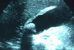
Ultrasound allows for evaluation of all the abdominal structures. It provides anatomic information about gallbladder size, stone size, gallbladder wall, and bile duct size.
Scintigraphy with hepatobiliary iminodiacetic acid (HIDA) scan can be considered if ultrasound results and CT results are equivocal, but it is rarely rapidly available in an acute situation.[40]American College of Radiology; American College of Nuclear Medicine; Society of Nuclear Medicine and Molecular Imaging; Society for Pediatric Radiology. ACR-ACNM-SNMMI-SPR practice parameter for the performance of hepatobiliary scintigraphy. 2021 [internet publication].
https://www.acr.org/-/media/ACR/Files/Practice-Parameters/hepato-scint.pdf
However, HIDA scan has the highest sensitivity and specificity for the diagnosis of acute calculus cholecystitis (ACC) as compared with other imaging modalities.[36]Pisano M, Allievi N, Gurusamy K, et al. 2020 World Society of Emergency Surgery updated guidelines for the diagnosis and treatment of acute calculus cholecystitis. World J Emerg Surg. 2020 Nov 5;15(1):61.
https://www.ncbi.nlm.nih.gov/pmc/articles/PMC7643471
http://www.ncbi.nlm.nih.gov/pubmed/33153472?tool=bestpractice.com
[37]Bonomo RA, Chow AW, Edwards MS, et al. 2024 clinical practice guideline update by the Infectious Diseases Society of America on complicated intra-abdominal infections: risk assessment, diagnostic imaging, and microbiological evaluation in adults, children, and pregnant people. Clin Infect Dis. 2024 Jul 5:ciae346.
https://academic.oup.com/cid/advance-article/doi/10.1093/cid/ciae346/7706348
http://www.ncbi.nlm.nih.gov/pubmed/38965057?tool=bestpractice.com
[38]Bonomo RA, Edwards MS, Abrahamian FM, et al. 2024 clinical practice guideline update by the Infectious Diseases Society of America on complicated intra-abdominal infections: diagnostic imaging of suspected acute cholecystitis and acute cholangitis in adults, children, and pregnant people. Clin Infect Dis. 2024 Jul 4:ciae349.
https://academic.oup.com/cid/advance-article/doi/10.1093/cid/ciae349/7706134
http://www.ncbi.nlm.nih.gov/pubmed/38963820?tool=bestpractice.com
Abdominal CT scan is inferior to ultrasound and magnetic resonance imaging (MRI) in assessing acute biliary disease, but it is useful when obesity or gaseous distension limits ultrasound interpretation. It is also indicated for evaluation of suspected complications (e.g., abscess) and concurrent intra-abdominal conditions. Abdominal MRI is appropriate for pregnant patients with abdominal pain and is recommended alongside abdominal ultrasound.[36]Pisano M, Allievi N, Gurusamy K, et al. 2020 World Society of Emergency Surgery updated guidelines for the diagnosis and treatment of acute calculus cholecystitis. World J Emerg Surg. 2020 Nov 5;15(1):61.
https://www.ncbi.nlm.nih.gov/pmc/articles/PMC7643471
http://www.ncbi.nlm.nih.gov/pubmed/33153472?tool=bestpractice.com
[37]Bonomo RA, Chow AW, Edwards MS, et al. 2024 clinical practice guideline update by the Infectious Diseases Society of America on complicated intra-abdominal infections: risk assessment, diagnostic imaging, and microbiological evaluation in adults, children, and pregnant people. Clin Infect Dis. 2024 Jul 5:ciae346.
https://academic.oup.com/cid/advance-article/doi/10.1093/cid/ciae346/7706348
http://www.ncbi.nlm.nih.gov/pubmed/38965057?tool=bestpractice.com
[38]Bonomo RA, Edwards MS, Abrahamian FM, et al. 2024 clinical practice guideline update by the Infectious Diseases Society of America on complicated intra-abdominal infections: diagnostic imaging of suspected acute cholecystitis and acute cholangitis in adults, children, and pregnant people. Clin Infect Dis. 2024 Jul 4:ciae349.
https://academic.oup.com/cid/advance-article/doi/10.1093/cid/ciae349/7706134
http://www.ncbi.nlm.nih.gov/pubmed/38963820?tool=bestpractice.com
[41]Watanabe Y, Nagayama M, Okumura A, et al. MR imaging of acute biliary disorders. Radiographics. 2007 Mar-Apr;27(2):477-95.
http://www.ncbi.nlm.nih.gov/pubmed/17374864?tool=bestpractice.com
Plain radiographs may detect a radiopaque gallstone in 15% of cases and provide information about bowel gas pattern or free air, but offer no incremental information if ultrasound or CT is performed.[42]Bortoff GA, Chen MY, Ott DJ, et al. Gallbladder stones: imaging and intervention. Radiographics. 2000 May-Jun;20(3):751-66.
http://www.ncbi.nlm.nih.gov/pubmed/10835126?tool=bestpractice.com
