Tests
1st tests to order
plain x-rays
Test
Ordered in all patients with a history of a fall or trauma who present with acute pain in the hip.[51]
Standard post-traumatic hip radiographs should include an anteroposterior (AP) view with 15° of internal hip rotation and a cross-table lateral view of the affected hip.[51][Figure caption and citation for the preceding image starts]: Initial anteroposterior radiograph showing a displaced left hip intracapsular fractureFrom the collection of Bradley A. Petrisor, MSc, MD, FRCSC and Mohit Bhandari, MD, MSc, FRCSC [Citation ends].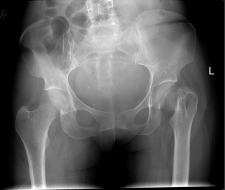 [Figure caption and citation for the preceding image starts]: Anteroposterior x-ray showing femoral neck fractureFrom the collection of Bradley A. Petrisor, MSc, MD, FRCSC and Mohit Bhandari, MD, MSc, FRCSC [Citation ends].
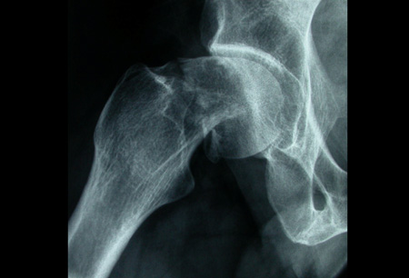
An x-ray of the femur can be ordered if distal extension of the fracture is suspected. Many institutions include an AP view of the pelvis for assessment of hip symmetry, either as part of the standard trauma evaluation or in conjunction with the dedicated hip series.[51]
Intracapsular fracture patterns: disruption of the cortex as well as the primary compressive and tensile trabecular lines suggests displacement in the fracture; increased angulation of varus or valgus on the AP image or increased anterior or posterior version as seen on the lateral radiograph also suggests displacement and can be compared with the other intact hip.
Extracapsular fracture patterns: stability of displaced fractures generally takes into account the extent of comminution and, more specifically, the comminution of the medial cortex. Simple 2-part intertrochanteric fracture patterns with no comminution of the medial calcar (cortex) are generally considered stable, and 3- and 4-part intertrochanteric fractures with disruption of the posteromedial cortex or the reverse-obliquity fracture are considered unstable.[3][52][Figure caption and citation for the preceding image starts]: Unstable intertrochanteric fracture on x-rayFrom the collection of Bradley A. Petrisor, MSc, MD, FRCSC and Mohit Bhandari, MD, MSc, FRCSC [Citation ends].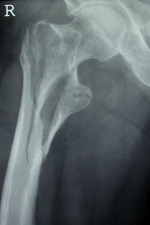
Result
may show fracture of proximal femur
Tests to consider
MRI pelvis (without contrast)
Test
Ordered if clinical suspicion of hip fracture is high with a negative plain x-ray.[51]
MRI has a higher sensitivity than plain radiographs for the detection of occult hip fractures.[51][54][55][Figure caption and citation for the preceding image starts]: MRI showing coronal imaging confirming an intracapsular fracture of the left hipFrom the collection of Bradley A. Petrisor, MSc, MD, FRCSC and Mohit Bhandari, MD, MSc, FRCSC [Citation ends].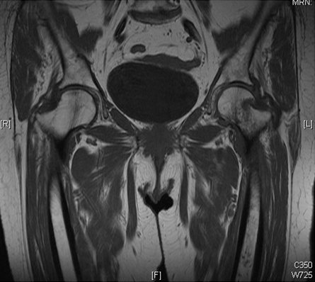
Result
presence of marrow edema and a fracture line
CT pelvis (without contrast)
Test
May be used if there is no access to an MRI.
Used to confirm suspicious finding on a plain x-ray or to confirm a fracture when clinical findings suggest a fracture but plain x-rays are negative.[51]
Result
may show fracture of proximal femur
Use of this content is subject to our disclaimer