Recommendations
Key Recommendations
Shock is often first suspected by low or declining blood pressure and acute deterioration in other vital signs, such as an increase in heart rate, increased respiratory rate, decrease in oxygen saturations, reduced level of consciousness, or decreased urinary output.
Shock may also be anticipated in relation to an established diagnosis, most typically acute myocardial infarction, severe infection, or severe hemorrhage. A patient in shock should be managed in a critical-care setting wherever possible.
Recognizing the patient in shock
Patients in shock look ill. They often describe symptoms specific to the underlying etiology (e.g., chest pain, shortness of breath, or abdominal pain). Altered cognition is the most sensitive and universal sign of hypoperfusion. Agitation and distress characterize early and milder stages, whereas unresponsiveness indicates more severe and advanced shock.[3]
Clinical signs of shock
Sustained hypoperfusion induces variable organ dysfunction, with signs dependent on the degree of hypoperfusion, duration of episode, and ability of the patient to compensate.
The most common signs include:
Stress responses (tachycardia, tachypnea)
Centralization of blood by peripheral vasoconstriction (cool extremities, mottled or ashen skin, slow capillary refill)[1][2][3]
Hypotension is usually defined as an adult systolic arterial blood pressure <90 mmHg, or a mean arterial pressure of <65 mmHg in septic shock (sepsis with hypotension).[2][25] A low diastolic blood pressure suggests arterial vasodilation (such as in anaphylaxis or sepsis).[26] Direct measurement via automatic cuff devices or an arterial line is preferred over routine sphygmomanometry, because direct blood pressure measurement is more precise and allows for continous monitoring. If an arterial line is used, this provides access for arterial blood sampling.[1] However, blood pressure is only an indirect measure of perfusion. A normal blood pressure reading does not rule out shock.[1] Hypoperfusion may be present at higher blood pressures. Sustained or severe hypoperfusion consistently leads to low blood pressure.
Narrowed pulse pressure (the difference between systolic and diastolic pressures; normally 35-45 mmHg) suggests arterial vasoconstriction (such as occurs in cardiogenic shock or hypovolemia) and may occur with rapid tachyarrhythmias.[26] However, pulse pressure is a function of both cardiac output and arterial function. As cardiac output is affected by the phase of respiration, pulse pressure should be measured at the same point in the respiratory cycle each time to enable comparison.
Hypotension in conjunction with signs of hypoperfusion is diagnostic of shock, and appropriate treatment should be instigated. By contrast, hypotension alone, without organ dysfunction, does not require immediate intervention, although regular review and monitoring are important.
The most useful parameters include change in cognition and deterioration in vital signs (heart rate, respiratory rate, temperature, and oxygen saturations). Capillary refill time may be delayed, indicating low skin perfusion, but it is not diagnostic of shock as it may be affected by age and environmental temperature.
Heart rate and blood pressure values can be used to calculate the Shock Index (SI). SI has been shown to be a clinically useful bedside method to estimate the level of shock and mortality in potential hemorrhagic and infection-related shock states. SI is determined by dividing the heart rate by systolic blood pressure. The normal range is 0.5 to 0.7; elevated SI >1.0 is associated with a need for critical care management and potential mortality.[27]
Determining the etiology of shock: history
While all causes of shock require prompt diagnosis and management of the underlying etiology, some require immediate treatment and must be readily identified (e.g., hemorrhage, tension pneumothorax, cardiac tamponade, arrhythmias, and anaphylaxis).
The history often reveals valuable information to narrow down the possible causes, but treatment is a priority in a patient with shock. In particular, ask about previous cardiac disease, medications (especially those recently started or stopped), any existing allergies and possible exposure to allergens, existing conditions such as abdominal aortic aneurysm, and any recent illness or hospitalization (which may increase the risk of sepsis or pulmonary embolism).
Determining the etiology of shock: examination
Rapid assessment and treatment usually occur concurrently. Cognition, pulse rate, blood pressure, and respiratory rate will reveal if a patient is improving or deteriorating with treatment.
Key findings on physical exam are:
Obstructive shock: tension pneumothorax is characterized by jugular vein distention, differences in chest excursion, resonance to percussion, decreased vocal fremitus, and absent breath sounds in the area of the pneumothorax, as well as tracheal deviation. If strongly suspected, immediate decompression should be performed by inserting an intravascular catheter in the second intercostal space in the midclavicular line (or the fifth intercostal space of the anterior axillary line) on the side of the pneumothorax and listening for the hiss of air escaping. Improvement in vital signs should follow. Jugular vein distention in combination with muffled heart sounds and bradycardia are characteristic of cardiac tamponade. Severe pulmonary embolism most commonly presents with increased respiratory rate (>20 breaths/minute), hypoxia, and a sinus tachycardia; these symptoms may be difficult to differentiate clinically from other causes of shock.
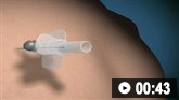 Needle decompression of tension pneumothorax: animated demonstration
How to decompress a tension pneumothorax. Demonstrates insertion of a large-bore intravenous catheter into the fourth intercostal space in an adult.
Cardiogenic shock: acute myocardial infarction is the most common cause of cardiogenic shock, but clinical signs are usually nonspecific.[4] Hypotension (systolic blood pressure <90mm Hg) is the primary clinical manifestation of shock but is not sufficient for the diagnosis.[4]
A new or changed murmur may be heard on cardiac auscultation if a valve abnormality is present. Typically, pulmonary edema occurs in severe cases with rales on lung auscultation and jugular vein distention; the skin is often mottled but may be moist with diaphoresis. Cardiogenic shock due to right ventricular failure may present with profound hypotension as a primary finding.
Distributive shock: spinal shock is usually indicated by an initially flaccid paralysis below the level of the lesion, with a history compatible with spinal trauma or a spinal lesion. The bladder may be palpated due to urinary retention. A key feature of anaphylaxis may be rapid facial and tongue swelling with airway control an urgent consideration. A widespread, itchy, erythematous rash, sweating, and warm peripheries are common. If anaphylaxis is likely, intramuscular epinephrine (adrenaline) is warranted. Septic shock requires evidence of infection and hypotension despite adequate cardiac output and filling. Signs may include either fever or low body temperature, warm peripheries, and signs specific to the site of infection. A petechial rash is characteristic for meningococcal septicemia, but may occur also with other microbes.
Hypovolemic shock: bleeding may be obvious (e.g., hematemesis, hematochezia, melena), or suggested by signs of severe trauma including long bone fractures. A tender abdomen may be present with intra-abdominal bleeding (e.g., ectopic pregnancy or ruptured abdominal aortic aneurysm). Extensive burns can account for considerable fluid losses. Septic shock also has qualities of hypovolemic shock.
Urgent investigations
Initial investigations are to diagnose shock, monitor the effect of interventions, and narrow down the potential etiologies. These are likely to include:
ECG to look for signs of cardiac ischemia (ST segment deviation, abnormal Q waves or loss of R wave, discordantly negative T waves or bundle branch block) and potentially critical arrhythmias, especially broad complex tachycardia.[3][15][31]
Complete blood count, serum chemistries, C-reactive protein, and lactate for evidence of sepsis, acute kidney injury, and tissue hypoperfusion; blood cultures should be taken when sepsis is suspected; elevated procalcitonin levels may help differentiate sepsis from causes of the systemic inflammatory response syndrome.[1][2][3][32]
Focused ultrasound of the abdomen and chest to demonstrate or exclude critical bleeding (e.g., tamponade, free abdominal fluid, or aortic aneurysm), evaluate cardiac function and cardiac cavity enlargement (particularly of the right ventricle in massive pulmonary embolism), assess for pneumothorax and pulmonary consolidation, and assess the diameter and filling of the inferior vena cava to determine vascular volume status.
Arterial blood gas (ABG) or venous blood gas (VBG) to assess acid-base status and oxygenation is useful in all causes of shock. The base deficit may help guide fluid resuscitation.[33] VBG is increasingly used as it is less invasive and less painful than ABG.[34]
Blood glucose for evidence of hypoglycemia or hyperglycemia, which may be a cause or a consequence of shock (e.g., diabetic ketoacidosis).[35]
Rapid urine pregnancy testing for women of childbearing age and pelvic ultrasound if a ruptured ectopic pregnancy is suspected.
Chest x-ray may offer clues to the etiology, such as a widened mediastinum in aortic disruption, hemothorax, or a globular cardiac shadow suggestive (but not diagnostic) of cardiac tamponade. Pneumonia can usually be confirmed by chest x-ray, but in early pneumonia or hypovolemic states there is potential that an existing pneumonia will not be detected with conventional x-ray.
Further investigations
Additional tests can be used to identify precise etiology and may include (depending on the likely etiology):
Ultrasound or computed tomographic (CT) scanning
Ultrasound can be performed at the bedside, whereas a patient must be stabilized before attempting a transfer to a CT scanner.
Echocardiography
This can be performed at the bedside
Echocardiography provides hemodynamic assessment, including cardiac output, stroke volume, ventricular filling, left ventricular afterload, and other parameters that help in determining the etiology of the shock state and aid in ongoing patient management.[1]
End tidal carbon-dioxide (capnography)
Side stream or direct capnography (when endotracheal airway is in place) can be helpful in assessing pulmonary perfusion matched to ventilation. In the critical care setting, below expected (or decreasing) end tidal carbon dioxide measured by capnography is associated with impaired matching of pulmonary perfusion and ventilation. This may assist in determining the presence of shock.[36]
Central venous catheter
A central venous line can be helpful, but requires time, special equipment, and skills, as well as a certain degree of patient stability. Central venous blood pressure and venous oxygen saturations may aid in differentiating the shock etiology, and judging treatment efficacy.[37] One randomized controlled trial reported that use of antibiotic- or heparin-impregnated central venous catheters reduced secondary (iatrogenic) blood stream infections.[38]

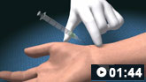
How to take a venous blood sample from the antecubital fossa using a vacuum needle.
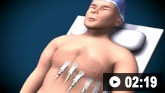
How to record an ECG. Demonstrates placement of chest and limb electrodes.
How to obtain an arterial blood sample from the radial artery.
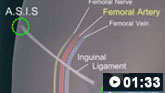
How to perform a femoral artery puncture to collect a sample of arterial blood.
Use of this content is subject to our disclaimer