Investigations
1st investigations to order
clavicle x-ray series
Test
Order for all patients with suspected clavicle fracture.[25][32] A standard clavicle x-ray series includes anteroposterior (AP) views in internal and external rotation and an axillary or scapular-Y view.[32] The exact angle used for the cephalic tilt view depends on patient positioning and body habitus to ensure the clavicle is projected clearly above the scapula and 2nd and 3rd ribs. Position the patient upright, rather than supine, as this may better demonstrate the degree of any displacement in a midshaft fracture.[35]
[Figure caption and citation for the preceding image starts]: Anteroposterior radiograph of right shoulder demonstrating clavicle fractureArnold S et al. BMJ Case Reports CP 2021;14:e241382; used with permission [Citation ends].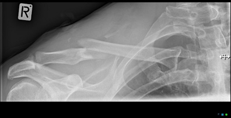 [Figure caption and citation for the preceding image starts]: Anteroposterior radiograph of left shoulder demonstrating a clavicle fractureAlao D et al. Emergency Medicine Journal 2005;22:232-3; used with permission [Citation ends].
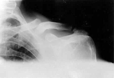
Result
fracture of clavicle, may also show acromioclavicular or sternoclavicular joint injury
CT chest, abdomen, pelvis
Investigations to consider
chest x-ray
Test
Order if suspected shortening of clavicle fracture (to enable comparison with opposite side) and/or suspected associated rib fracture. A posteroanterior view should be taken.
Result
fracture of clavicle and/or rib
scapula x-ray series
Test
Order if suspected scapula fracture.
Result
fracture of scapula
shoulder x-ray series
Test
Order if suspected injury to the proximal humerus or glenohumeral joint. AP view in internal and external rotation and an axillary or scapular-Y view should be taken.[32] Axillary or scapula-Y views are vital in evaluating traumatic shoulder injuries as acromioclavicular and glenohumeral joint dislocations can be misclassified on AP views.[32][33][34]
Result
fracture of clavicle and/or fracture of humerus, dislocation of glenohumeral join
CT clavicle
Test
Reserved for cases in which the clinician suspects a more complex injury than the plain radiographs show (such as a sternoclavicular joint dislocation).[32][38] A CT will give an accurate estimate of shortening and displacement, and may impact treatment planning.
Result
fracture of clavicle; provides more accurate estimate of shortening and displacement than clavicle x-ray
ultrasound of clavicle
Test
Ultrasound is rarely indicated in the evaluation of clavicle fractures.[32] May be useful, however, for detection of fracture-associated haematoma, and can show fracture at surface of bone if present and/or joint instability. Request ultrasound if there is a high index of suspicion for clavicle fracture but the injuries are not seen on plain x-rays, or if trying to avoid radiation exposure (e.g., in a child or a pregnant patient).[39]
Result
fracture of clavicle and/or dislocation at sternoclavicular or acromioclavicular joint
MRI of acromioclavicular or sternoclavicular joint
Use of this content is subject to our disclaimer