Differentials
Testicular torsion
SIGNS / SYMPTOMS
Typical history of sudden onset of testicular pain.
Physical examination shows a horizontal, rotated, high-riding testis, painful to touch. This is in contrast to a low testis in standing position characteristic of testicular mass.
Neonates typically present with a unilateral swollen and discoloured scrotum of sudden onset.
INVESTIGATIONS
Ultrasound shows a diffuse hypoechogenicity and enlargement when compared with the contralateral side. However, the testis may appear normal in the first 6 hours. In these cases a rotated spermatic cord on examination can be a highly reliable and direct sign. Doppler ultrasound will detect blood flow occlusion in most cases after the first 6 hours. Heterogenicity after 24 hours indicates infarction and haemorrhage. Markers are negative.
Epididymo-orchitis
SIGNS / SYMPTOMS
Signs of inflammation (erythema, warmth, and pain) may occur and are not usually seen with malignancy.
Clinical history is usually shorter (<2 weeks).
INVESTIGATIONS
Colour Doppler imaging characteristically shows increased blood flow to the testicle and epididymis. Occasionally an abscess can be detected in immunocompromised patients. If there is doubt, tumour markers may be checked, which should be negative.
Diagnosis by urethral swab and culture can detect associated STDs.[58]
Chronic orchitis may present as a shrunken testis with increased echogenicity on ultrasound. Thickening of the capsule and fluid collections can be seen surrounding the testis.
Scrotal hernia
SIGNS / SYMPTOMS
Bowel sounds may be heard on examination. If the mass can be reduced on palpation this indicates a hernia.
INVESTIGATIONS
A characteristic scrotal ultrasound finding is herniated bowel in the scrotum. The bowel herniates through a patent processus vaginalis and is separate from the testis. Loops of bowel with peristaltic movements and omentum can be seen.
Hydrocele
SIGNS / SYMPTOMS
On physical examination the mass transilluminates with light. The fluid collection usually engulfs the whole testis so the testis cannot be palpated. However, a testicular mass can coexist with hydrocele and should be ruled out by scrotal ultrasound.
INVESTIGATIONS
High-resolution ultrasound will differentiate from intratesticular solid mass. The mass is translucent and surrounds the whole testis.
Epididymal cyst
SIGNS / SYMPTOMS
Scrotal mass that can be transilluminated. The position of the cyst helps the diagnosis. The cyst can be palpated separately from the testis.[59]
INVESTIGATIONS
Scrotal ultrasound may assist in making the diagnosis.
Haematomas
SIGNS / SYMPTOMS
There may be a recent history of blunt trauma. However, up to 10% of testicular cancer also presents after a history of trauma.[42]
INVESTIGATIONS
There are no specific tests. Diagnosis is made based on history of trauma and clinical examination.
Ultrasound may show a break in the tunica albuginea, testicular contour irregularity, and poorly defined borders of the testicle.[Figure caption and citation for the preceding image starts]: Longitudinal grey-scale image of the medial right testicle shows a 5.4×5.6 cm intratesticular heterogenous focus with a peripheral hypoechoic rimBMJ Case Reports 2010; doi:10.1136/bcr.06.2010.3119 [Citation ends].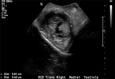 Serum tumour markers are negative.
Spermatocele
SIGNS / SYMPTOMS
On examination this should be separate from the testis and near the high pole. Easily distinguished using a scrotal ultrasound.
INVESTIGATIONS
Ultrasound is reliable in visualising a small fluid-filled cyst.
Intratesticular benign cysts
SIGNS / SYMPTOMS
Most are not palpable except when they are at the periphery of the testis. These include intratesticular simple cysts, tubular ectasia, epidermoid cyst, tunica albuginea cyst, and intratesticular varicocele.
INVESTIGATIONS
Ultrasound will detect a fluid-filled cavity within the testis in almost 100% of the cases.[45]
Syphilitic gumma
SIGNS / SYMPTOMS
Inflammatory, fibrous nodules that may cause local destruction. Can occur anywhere but most often affect the bone and skin.
Gumma are a late manifestation of syphilis. May have a history of secondary syphilis. Signs of early syphilis include general lymphadenopathy and genital and mucosal ulceration. Signs of late syphilis include pupillary changes (Argyll-Robertson pupil) and aortic regurgitation.[60]
INVESTIGATIONS
Positive treponema pallidum serology.
Use of this content is subject to our disclaimer