Aetiology
Rickettsial infections occur worldwide and are associated with the patient having been bitten by an ectoparasite such as a louse, mite, flea, mosquito, or tick.[18][19][20] Around 200 vector species have been detected, carrying 47 Rickettsia spp, with ticks big the most common vector.[7] These small arthropods contain the rickettsiae living inside them, usually as part of their normal microbial flora, and they are transmitted to the patient by the bite of the ectoparasite or via its infected faeces, either when rubbed into the bite site or when the ectoparasite faeces are inhaled by the patient. CDC: tick ID Opens in new window
Rickettsial infections in humans are caused by several related genera of bacteria including Rickettsia species, Orientia tsutsugamushi and O chuto, Anaplasma species, Ehrlichia species, and Neoehrlichia species. Sometimes Coxiella burnetii (which causes Q fever) is included as a rickettsiosis, but strictly speaking it is not as it belongs to a phylogenetically different clade of bacteria and is more closely related to Legionella species. See the Classification section for a list of types of rickettsioses, causative organisms, and vectors.
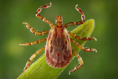
Rickettsia have two common features: they are obligate intracellular bacteria and will only grow inside a host animal eukaryotic cell, and they live in the cells of both vertebrate (e.g., humans, rats) and invertebrate animals (e.g., lice, fleas, mites, and ticks). While the invertebrate is the main host animal for the rickettsia, it is also the vector for transmission of the rickettsia to humans and other vertebrate animals.[Figure caption and citation for the preceding image starts]: Male yellow dog tick (Amblyomma aureolatum)CDC/Dr Christopher Paddock and James Gathany (photographer) [Citation ends].
Pathophysiology
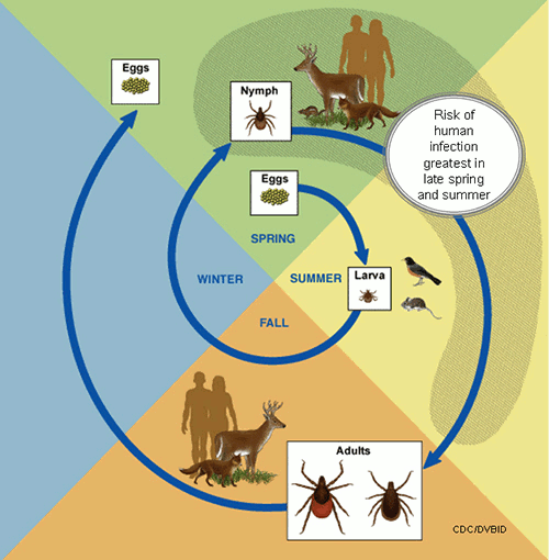
Rickettsia are taken up by human cells that are not professional phagocytes. The cell type most favoured is the endothelial cell, leading to leaking tight junctions, oedema, capillary destruction, and petechiae. This pathology can occur in any end organ and lead to thrombosis, infarction, and tissue necrosis. There is some variation between the different rickettsial pathogens, but it is variation on a theme. Brain, lung, spleen, liver, and kidney may all be damaged by the intracellular growth of the rickettsia. There is not thought to be a rickettsial toxin involved, but simply the destruction of the patient's endothelial cells by the intracellular growth of the rickettsiae.[21][Figure caption and citation for the preceding image starts]: Life cycle of ticksCDC [Citation ends].
Classification
Centers for Disease Control and Prevention classification[2]
Scrub typhus group rickettsioses
Due to mite-transmitted infection with the rickettsia Orientia tsutsugamushi, O chuto, or O chiloensis.
Typhus group rickettsioses
Epidemic typhus, sylvatic typhus: due to infection by human body lice themselves infected with Rickettsia prowazekii. Now rare, but may occur in situations of mass crowding, poverty, famine, and war.
Murine typhus: due to infection with R typhi from the infectious faeces of rodent fleas, living on rodents in close proximity to humans.
Spotted fever group rickettsioses
Different species occur in geographically different parts of the world. About 20 different infections are classified in this group, including:
African tick-bite fever (tick transmitted) due to R africae
Aneruptive fever (tick transmitted) due to R helvetica
Astrakhan spotted fever (tick transmitted) due to R conorii (subspecies caspiae - proposed)
Cat-flea rickettsiosis (flea transmitted) due to R felis
Far Eastern spotted fever (tick transmitted) due to R heilong-jiangensis
Flinders Island spotted fever or Thai tick typhus (tick transmitted) due to R honei (including marmionii strain)
Indian tick typhus (tick transmitted) due to R conorii (subspecies indica - proposed)
Israeli tick typhus (tick transmitted) due to R conorii (subspecies israelensis - proposed)
Japanese spotted fever (tick transmitted) due to R japonica
Lymphangitis-associated rickettsiosis (tick transmitted) due to R siberica mongolotimonae
Maculatum infection (tick transmitted) due to R parkeri
Mediterranean spotted fever or Boutonneuse fever (tick transmitted) due to R conorii (subspecies conorii - proposed)
Mediterranean spotted fever-like disease (tick transmitted) due to R massiliae or R monacensis
North Asian tick typhus or Siberian tick typhus (tick transmitted) due to R sibirica
Queensland tick typhus (tick transmitted) due to R australis
Rickettsialpox (mite transmitted) due to R akari
Rickettsiosis (tick transmitted) due to R aeschlimannii
Rocky Mountain spotted fever (tick transmitted) due to R rickettsii
Tickborne lymphadenopathy or Dermcentor-borne necrosis and lymphadenopathy (tick transmitted) due to R raoultii or R slovaca.
Use of this content is subject to our disclaimer