Investigations
1st investigations to order
mammography
Test
Patients who have a history of a persistent breast mass, are suspected to have a mass on breast examination, or have suspicious nipple discharge should undergo diagnostic mammography and breast ultrasound.[1][2][20]
Breast biopsy is indicated for solid masses on imaging studies to differentiate between fibrocystic breasts and malignancy.
Suspicious nipple discharge is most often caused by an intraductal papilloma (70%), breast cancer (5%), or ductal ectasia (25%).
Breast pain alone is not an indication for imaging in the absence of other physical findings suggestive of an anatomical cause for the pain, such as a cyst or mass.[16][17] Extensive workup of patients who present only with mastalgia is not indicated, other than age-appropriate screening mammography.
Result
dense breasts, circumscribed density
breast ultrasound
Test
Patients who have a history of a persistent breast mass, who are suspected to have a mass on breast examination, or who have suspicious nipple discharge should undergo diagnostic mammography and breast ultrasound.[1][2][20]
Cysts must be confirmed by breast ultrasound. Based on ultrasound criteria, cysts may be simple, indeterminate, or complex. Characteristically, simple cysts are anechoic, have smooth, sharp margins, and have retrotumoural acoustic enhancement. Complex cysts have a solid component, mural growth, wall thickening, or thick septa, and require diagnostic biopsy.
Breast biopsy is indicated for solid masses on imaging studies to differentiate between fibrocystic breasts and malignancy.
Suspicious nipple discharge is most often caused by an intraductal papilloma (70%), breast cancer (5%), or ductal ectasia (25%).
Breast pain alone is not an indication for imaging in the absence of other physical findings suggestive of an anatomical cause for the pain, such as a cyst or mass.[16][17] Extensive workup of patients who present only with mastalgia is not indicated, other than age-appropriate screening mammography.
[Figure caption and citation for the preceding image starts]: Ultrasound image of a breast cyst (note the characteristic smooth and sharp margins of the anechoic lesion with posterior acoustic enhancement)Courtesy of Limin Yang, MD, and Justin Boatsman, MD, Department of Radiology, University of Iowa Hospital and Clinics; used with permission [Citation ends].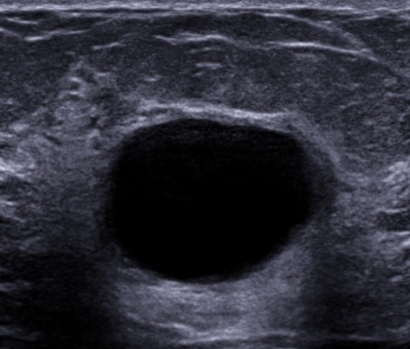
Result
breast cysts, solid mass
Investigations to consider
cyst aspiration
Test
Indicated for women who have symptomatic breast cysts.
If fluid aspirated is straw-coloured and the cyst is completely aspirated there is no need for cytological studies.
If the fluid aspirated is bloody, cytology of the fluid or biopsy of the cyst is recommended.[1][2][20]
Result
straw-coloured, bloody fluid
breast biopsy
Test
Indicated for solid masses on palpation or imaging studies.
Also indicated if there is a palpable mass in spite of negative imaging studies.
Histological findings that confirm fibrocystic breasts are apocrine metaplasia and hyperplasia, gross and microscopic cysts, and fibrosis.[1][2] Other alterations include certain stromal alterations, mild epithelial hyperplasia, and mild degrees of adenosis.
Primary role of biopsy is to exclude breast cancer or a high-risk pathology.[1][2][20]
Patients with suspicious nipple discharge and normal mammography and breast ultrasound still require a tissue diagnosis and an excisional biopsy of the secreting duct and underlying breast tissue (microductectomy or central duct excision).
Diagnostic biopsy, to exclude breast cancer or an intracystic papilloma, is indicated for cysts demonstrating sonographically complex characteristics, such as a solid component, mural growth, wall thickening, or thick septa.
Result
apocrine metaplasia, fibrosis, cyst formation, and proliferative changes, atypical ductal hyperplasia
Use of this content is subject to our disclaimer