Images and videos
Images


Eye trauma
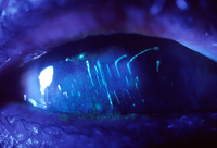
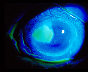
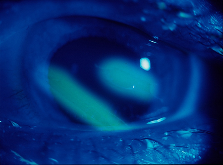
Corneal ulcer seen with fluorescein stain
Used with kind permission from Drs Smith, Severn, and Clarke
See this image in context in the following section/s:
Eye trauma
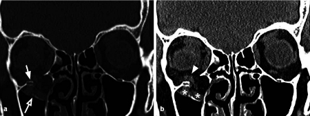
Coronal CT reconstruction with bone (a) and soft tissues (b) algorithms of an isolated orbital floor fracture. a Right orbital floor fracture with involvement of the medial aspect of the infraorbital groove (white arrow) and dislocation of a bony fragment into the maxillary sinus (empty arrow). b Mild swelling of the right inferior rectus muscle (arrowhead) partially herniated into the bony defect; huge herniation of the intraorbital fat through the fracture gap; within the herniated soft tissue, hematomas are recognizable (asterisks)
Cellina M et al. Insights Imaging 2022 Jan 12;13(1):4; used with permission
See this image in context in the following section/s:

Eye trauma
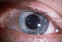
Corneal foreign body
Used with kind permission from Drs Smith, Severn, and Clarke
See this image in context in the following section/s:

Eye trauma
The ABCs of ocular trauma evaluation
Adapted from Kroesen CF et al. Mil Med 2020 Jan 7;185(suppl 1):448-53
See this image in context in the following section/s:
Eye trauma
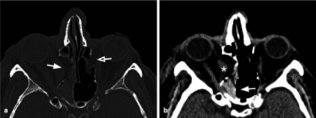
Axial CT acquisition with bone (a) and soft tissues (b) algorithms in a patient with a current fracture of the right orbital medial wall (white arrow) and a previous fracture of the contralateral orbital medial wall (empty arrow). a Extensive discontinuation of the right lamina papyracea (white arrow). b The medial rectus muscle is swollen, displaced medially, and partially entrapped into the fracture (asterisk); presence of hemosinus, visible as a right posterior ethmoidal cell occupied by hyperdense material (blood) (arrow)
Cellina M et al. Insights Imaging 2022 Jan 12;13(1):4; used with permission
See this image in context in the following section/s:

Eye trauma
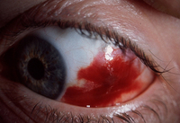
Sub-conjunctival haemorrhage
Used with kind permission from Drs Smith, Severn, and Clarke
See this image in context in the following section/s:

Eye trauma
Sub-tarsal foreign body. Vertical corneal abrasions
Used with kind permission from Drs Smith, Severn, and Clarke
See this image in context in the following section/s:
Eye trauma
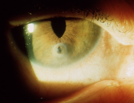
Penetrating corneal injury with iris prolapse
Used with kind permission from Drs Smith, Severn, and Clarke
See this image in context in the following section/s:
Eye trauma
Corneal abrasion seen with fluorescein stain
Used with kind permission from Drs Smith, Severn, and Clarke
See this image in context in the following section/s:
Use of this content is subject to our disclaimer