Approach
The aim of diagnosis is to identify patients who have an ongoing underlying cause for their fever.
Fever may be accompanied by other signs and symptoms. A systematic evaluation is paramount to guide diagnostic tests and establish the diagnosis. The differential diagnosis is largely influenced by the time elapsed between the surgical procedure and the onset of the fever.
History
The main aim of the history is to identify risk factors, known underlying conditions, and symptoms that suggest a cause other than an inflammatory response to surgery. The type of surgery performed, and the timing and duration of associated invasive catheterisation and intubation, should be noted.
Pain in the operative site and/or the wound is an important sign that may indicate infection, haematoma, or ischaemia of the operated tissue, especially if the pain is new or has become worse.
Drug history
It is important to identify and assess the anaesthetics, medications, and blood products used during and after the surgery, as many of these agents can cause a fever.
Malignant hyperthermia can present up to 1 hour after the administration of an inhalation anaesthetic or succinylcholine.[18] It is very rare.[18] The fever is usually >40ºC (104ºF) and associated with muscle rigidity, hypercapnia and/or tachypnoea, or tachycardia.
Blood or blood products may cause a transfusion reaction, which usually occurs during or immediately after the transfusion. Fever may be accompanied by headache, nausea, vomiting, anxiety and/or pain in the chest, back, abdomen or infused extremity. Temperature, heart rate, and blood pressure should be checked 15 minutes after the start of a blood transfusion, and the transfusion should be stopped immediately if a reaction is suspected.[29]
Beta-lactam antibiotics, cephalosporins, sulfonamides, vancomycin, rifampin, or fluoroquinolones can all cause fever, and may be used for perioperative antibiotic prophylaxis.[17] Other agents that can cause fever and are commonly given as part of the perioperative management include hydroxyurea, propylthiouracil, iodides, heparin, allopurinol, immunoglobulins, salicylates, phenytoin, hydralazine, procainamide, furosemide, and thiazide diuretics.[43]
Past medical history
The past medical history may include conditions that predispose the patient to developing a postoperative fever.
A history of alcohol abuse should raise suspicion of alcohol withdrawal.
Hyperthyroidism, underlying malignancy, and phaeochromocytoma predispose the patient to developing a non-infectious postoperative fever.
Conditions that produce immunosuppression or require immunosuppressive therapy predispose the patient to infection.
Patients with poor nutritional status have increased susceptibility to infection.
A thorough systems review should be undertaken to identify key associated symptoms of underlying causes.
Respiratory factors
Breathlessness with a productive cough may indicate pneumonia. Breathlessness with pleuritic chest pain and/or haemoptysis may be due to a pulmonary embolism (PE). The modified Wells and Geneva scores use risk factors and clinical features to suggest the probability of a PE; a high probability of PE should prompt diagnostic tests.
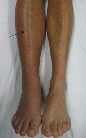
Leg swelling may indicate deep venous thrombosis (DVT). The probability of DVT can be assessed using the Wells clinical probability tool; a high probability of DVT should prompt diagnostic testing.
[Figure caption and citation for the preceding image starts]: Deep vein thrombosis (DVT) of the right leg with associated swelling and rednessFrom the collection of James Heilman, MD. [Citation ends].
Cardiovascular factors
Myocardial infarction typically presents with central chest pain that is heavy, crushing or ‘squeezing’ in nature. Pain may radiate to the left arm, neck or jaw and be associated with nausea, vomiting, diaphoresis, dyspnoea, lightheadedness, palpitations or syncope.
Pain can be atypical or absent in patients with diabetes.[44]
Gastrointestinal factors
New-onset abdominal pain may indicate a range of postoperative complications.
Peritonitis presents with severe abdominal pain and re-bound tenderness. If an underlying abscess is present, the location of the pain can vary, but is often associated with ileus, abdominal bloating, and a spiking fever.
Acalculous cholecystitis presents with severe right upper quadrant pain radiating to the right side. Pancreatitis presents with a mild epigastric pain radiating to the back.
Diarrhoea, especially if occurring after antibiotic use, may indicate Clostridium difficile infection.
Suprapubic pain may indicate a urinary tract infection (UTI). If the urinary catheter was recently removed, the patient may also report symptoms of dysuria, frequency, and urgency.
Neurological factors
The onset of new neurological symptoms indicates a serious underlying cause.
New-onset focal sensory or motor weakness may indicate an ischaemic or haemorrhagic stroke.
New-onset severe headache with photophobia may indicate a subarachnoid haemorrhage, meningitis, or subdural haematoma.
Rheumatological factors
Acute onset of joint pain and swelling may indicate an exacerbation of gout or pseudogout triggered by the stress of surgery.
In patients who have undergone orthopaedic surgery, osteomyelitis is a possibility.
Dermatological factors
Skin rashes occur with a range of underlying causes including drug reactions, fat embolism, meningitis, and toxic shock syndrome (TSS).
Inflammation of the skin or catheter insertion sites may be due to cellulitis or superficial thrombophlebitis.
A fluctuant mass may be a haematoma or seroma; seromas are usually painless.
[Figure caption and citation for the preceding image starts]: Rash and subcutaneous oedema of the right hand due to toxic shock syndromeFrom the CDC and the Public Health Image Library [Citation ends].

Ear, nose, and throat (ENT) factors
Some patients may have ENT symptoms. Nasal discharge with or without sinus pain suggests an acute sinusitis.
Parotid/submandibular pain on swallowing suggests sialadenitis and is particularly common in patients with poor oral hygiene, malnutrition, and older people.
Dysphagia may indicate stroke, and is also seen in sialadenitis.
Otalgia may indicate otitis media.
Organ transplant
Transplant rejection should be considered in patients who have had a recent organ transplant, although fever is an uncommon manifestation of rejection. Most patients report malaise, which is a non-specific symptom. Other symptoms are related to failure of the transplanted organ.
Kidney recipients may develop oliguria.
Lung recipients may report shortness of breath.
Liver recipients may report jaundice and bleeding.
Symptoms can be hyperacute (onset within minutes or hours) or acute (onset usually within weeks).
Examination
Physical examination should begin with a thorough evaluation of the vital signs, followed by a systematic examination, keeping in mind the differentials suspected on the basis of the surgical procedure performed and the time of onset of the fever.
General examination
The extent and pattern of fever should be noted. Tachypnoea, tachycardia, and hypotension are indications of a serious underlying cause. The general appearance of the patient reflects the seriousness of the underlying cause.
Respiratory distress or cyanosis indicates an underlying respiratory cause such as pneumonia or PE.
Decreased conscious state may indicate a neurological cause such as stroke or subarachnoid haemorrhage.
Wounds and foreign bodies
Inflammation, tenderness, or purulent drainage of a surgical wound indicates infection. Purulent wound drainage is seen with a range of superficial and deep wound infections.
A fluctuant mass could indicate a haematoma or seroma. Seromas are usually painless.
Inflammation, pain, tenderness, or purulent discharge of catheter insertion sites indicates superficial thrombophlebitis or cellulitis. Thrombophlebitis is particularly common with central venous catheters.
Cloudy urine may indicate a UTI.
Extremities
Examination of limbs may reveal leg swelling and calf tenderness, signs of a DVT. The presence of erythema, swelling, or rashes on the skin should be noted. Swelling, erythema, and tenderness of joints indicate gout, pseudogout, or osteomyelitis. Osteomyelitis results in decreased range of motion of the affected joint. A haematoma may produce a compartment syndrome, presenting with pain, pressure, and paraesthesia of the affected extremity; pallor and pulselessness are late signs.
Respiratory examination
Respiratory examination may reveal unilateral crepitations, poor air entry, or bronchial breathing, which indicate pneumonia.
Atelectasis is not a cause of fever, but is a common coincidental condition that may be detected as part of the respiratory examination.
Abdominal examination
Abdominal examination may reveal point tenderness, abdominal bloating, or abdominal abscess; peritonism may be noted. The appearance of these signs may indicate acalculous cholecystitis, pancreatitis, peritonitis (including peritonitis due to anastomotic breakdown), abdominal abscess, or ischaemia of the operated tissue.
Patients with cholecystitis may have a positive Murphy's sign.
A rectal examination may detect a pelvic abscess.
Neurological examination
Neurological examination may reveal focal sensory or motor weakness in patients with stroke, and neck stiffness with or without Kernig's sign in patients with meningitis.
Tremor may be an early sign of alcohol withdrawal.
Decreased conscious level may be produced by any bleeding or inflammation in the brain.
Cavernous sinus thrombosis may present with periorbital oedema, chemosis and proptosis, lateral gaze palsy and ophthalmoplegia.
ENT examination
ENT exam may reveal sinus tenderness in sinusitis, facial swelling over the parotid region in sialadenitis, or exudates of pus from salivary gland openings in the oral cavity in bacterial sialadenitis.
A compartment syndrome of the orbit may occur following surgery for facial trauma or eye surgery, and presents with eye pain, diplopia, visual loss, reduced ocular motility, and proptosis of the affected eye.
Bulging tympanic membrane may indicate otitis media.
Investigations
Initial testing is greatly influenced by the history and physical examination. Fever occurring in the first 48 hours is unlikely to be infectious and does not need further diagnostic testing unless there are associated signs or symptoms that strongly suggest an infection or other underlying cause, or the patient is immunocompromised.[45]
Immunocompromised patients require immediate thorough investigation for infection with a full blood count (FBC), chest radiograph, urinalysis with culture, and blood cultures, as these patients are vulnerable to opportunistic infections that can present at any time.
After 48 hours, a full blood count, chest radiograph, urinalysis with culture, blood cultures, and wound cultures are required as first tests in all patients. Further radiological or laboratory testing is directed toward the suspected aetiology.
A high white cell count and elevated C-reactive protein are useful indicators of infection, but are not specific.
Urine cultures identify urinary tract infections and blood cultures identify systemic bacterial infections from a wide range of sources, including wound infections, abscesses, catheter-related intravascular infections, foreign body infections, pneumonia, and transfusion-related infections. Wound cultures are helpful for surgical site infections, especially when there is associated purulent drainage. Sputum cultures are useful in patients with suspected pneumonia. A chest radiograph may show unilateral infiltration, consolidation, effusions, or cavitation consistent with pneumonia.
Drug-induced fever can present any time within 7 days of the drug being started, and all potential fever-inducing agents should be discontinued; if the fever resolves, a diagnosis of drug-induced fever can be made retrospectively.
Further tests are determined by the likely diagnosis.
Suspected MI: ECG should be performed and serum cardiac enzymes measured.
Suspected venous thromboembolism: the clinical suspicion of DVT can be graded using the Wells clinical probability score. [ Modified Wells score for deep vein thrombosis (DVT) Opens in new window ] Patients with a high probability of DVT require further investigation with a duplex ultrasound of the lower limb. The modified Wells or Geneva scores grade the clinical suspicion of a pulmonary embolus. [ Pulmonary Embolism Wells Score Opens in new window ] Patients with a high probability of pulmonary embolus require further investigation with computed tomography (CT) pulmonary angiography or ventilation-perfusion scanning (for patients who have a contraindication to CT pulmonary angiogram).
Stroke, subarachnoid hemorrhage, and subdural haematoma: can be diagnosed by a CT scan of the head. This should be performed in any patient with a postoperative fever and new-onset focal neurological signs or severe headache.
Ischaemia or infarction of the operated tissue: arterial blood gases reveal acidosis and elevated lactate.
Fat embolism: should be suspected in patients with recent major trauma or orthopedic surgery presenting with fever within 48 to 72 hours postoperatively. Chest x-ray may reveal a diffuse interstitial pattern typical of fat embolism, and CT scanning of the chest reveals a ground glass appearance of the lungs.[46][47]
C difficile infection: stool samples for C.difficile toxins in any patient with diarrhoea.
Wound seromas and haematomas: can be distinguished by ultrasound. Hypoechoic fluid collection and associated pain indicates haematoma; anechoic fluid indicates seroma. Seromas are typically painless.
Compartment syndromes: can be diagnosed by measurement of pressure in the affected compartment.
Suspected acalculous cholecystitis: abdominal ultrasound scan typically shows a thickened emphysematous gallbladder in the absence of stones.
Pancreatitis: serum lipase or amylase should be measured if pancreatitis is suspected. Use serum lipase testing in preference to serum amylase.[48][49][50] [
 ]
]
Blood transfusion reaction can be confirmed by inspection of centrifuged plasma and urine; the transfusion reaction produces haemoglobinemia, producing a clear red colour in centrifuged plasma and urine.
Suspected malignant hyperthermia: inhalation anaesthetics should be discontinued and dantrolene administered; resolution of symptoms with dantrolene supports the diagnosis, but confirmation of the diagnosis involves specialised tests at a later date.
Suspected gout or pseudogout: joint aspiration. The aspirated synovial fluid should be sent for Gram stain, microbial culture and polarised light microscopy to detect crystals.
Suspected endocrine cause: serum thyroid stimulating hormone, triiodothyronine and thyroxine should be checked in patients with hyperthyroidism; patients with a known pheochromocytoma require a measurement of serum-free metanephrines (metadrenalines) and normetanephrines (normetadrenalines) and a 24-hour urine collection for catecholamines, metanephrines (metarenalines), normetanephrines (normetadrenalines), and creatinine; serum cortisol and adrenocorticotrophic hormone should be measured in suspected adrenal crisis.
Hyperacute transplant rejection: may present minutes to hours after the transplant, but is exceedingly rare; a tissue biopsy is required to confirm the diagnosis.
Patients in intensive care who have recently undergone thoracic, abdominal, or pelvic surgery, may require a CT scan if the aetiology is not apparent from initial investigations.[51] A bedside abdominal ultrasound scan is recommended if there are abdominal symptoms or suspicion of an abdominal source of the fever, in intensive care patients who have had recent abdominal surgery.[51]
Investigations for specific surgical site infections are as follows
Deep abscesses: of which the most common are abdominal abscesses, are diagnosed by CT scanning of the affected compartment.
Suspected meningitis: requires a lumbar puncture to establish the diagnosis and detect infection. Most cases of meningitis after neurosurgery are aseptic, but infectious causes must be excluded.
Cavernous sinus thrombosis: a serious complication of head and neck surgery or neurosurgery. It is triggered by a remote infection, and cultures of blood and suppurative fluid or tissue from the primary infectious source are required. The diagnosis is made by contrast-enhanced high-resolution CT scanning of the head.
Foreign body infections: usually diagnosed based on blood cultures and cultures of wound discharge, but can be confirmed using x-rays of joint prostheses and technetium-methylene diphosphonate scintigraphy with leukocyte scanning. Synovial fluid culture and tissue culture should also be performed when joint prosthesis infection is suspected.[20]
Osteomyelitis: erythrocyte sedimentation rate, C-reactive protein, plain radiographs and magnetic resonance imaging can be helpful, but either bone biopsy or blood cultures will confirm the diagnosis and guide appropriate antibiotic selection.
Sinusitis and sialadenitis: may be triggered by intubation and the use of anaesthetics. Sialadenitis is diagnosed based on a combination of clinical features and the presence of sialoliths on facial radiographs or CT scanning. Culture of exudates from the duct is required to identify the causative organism. Sinusitis is a clinical diagnosis.
Infectious endocarditis following valve surgery: diagnosed by blood cultures and echocardiography. Echocardiography may reveal valvular vegetations. 3 sets of blood cultures should be obtained 1 hour apart before initiating empirical antibiotic therapy.
Use of this content is subject to our disclaimer