This page compiles our content related to dermatitis. For further information on diagnosis and treatment, follow the links below to our full BMJ Best Practice topics on the relevant conditions and symptoms.
Introduction
Relevant conditions
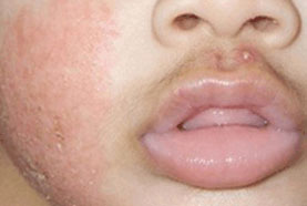
EczemaSigns & symptomsInvestigationsDifferentialsTreatment algorithm | go to our full topic on Eczema Eczema (atopic dermatitis) is characterised by dry, pruritic skin with a chronic relapsing course. One systematic review reported 12-month eczema prevalence of 1.8% to 17.0% in children in Europe, and 0.94% to 22.6% in children in Asia.[1] Eczema has a multifactorial aetiology, with a combination of genetic susceptibility and environmental factors contributing to disease development. Typically there is erythema, scaling, vesicles, or lichenification in skin flexures. In infants, the extensor surfaces, cheeks, scalp, and forehead are preferentially affected.[2] Patients often have a personal or family history of other atopic diseases such as asthma or allergic rhinitis.[2][3][4] Can be described as acute or chronic. Acute eczema is used to describe a flare-up of symptoms. Chronic eczema is used to describe the condition when the patient develops signs of chronic inflammation (e.g., lichenification). Diagnosis is primarily clinical.[Figure caption and citation for the preceding image starts]: Acute atopic dermatitis on the face of an infantPersonal collection of Dr A. Hebert [Citation ends]. |
|---|---|
Contact dermatitisSigns & symptomsInvestigationsDifferentialsTreatment algorithm | go to our full topic on Contact dermatitis Irritant contact dermatitis (ICD) is caused by direct toxicity and can occur in any person without prior sensitisation, whereas allergic contact dermatitis is a delayed hypersensitivity reaction. In one meta-analysis of 28 studies of over 20,000 individuals, the worldwide prevalence of contact dermatitis in the general population is estimated to be 20.1%.[5] ICD is the most common form of contact dermatitis and is due to direct, cytotoxic barrier damage. Common causes of ICD include chronic exposure to mild irritants (water, soaps, solvents, cutting fluids) or from acute exposure to more toxic agents (acids, alkalis, strong oxidising or reducing agents, organic solvents, and gasses). Patients generally report pruritus, burning, erythema, swelling, and blistering with acute contact dermatitis, and pruritus, burning, erythema or hyperpigmentation, fissuring, and scaling with chronic contact dermatitis. The clinical presentation of contact dermatitis is highly variable, depending on the causative agent, the affected body areas, and the duration of symptoms. |
Dyshidrotic dermatitisSigns & symptomsInvestigationsDifferentialsTreatment algorithm | go to our full topic on Dyshidrotic dermatitis A form of chronic dermatitis affecting the hands and feet. Characterised by recurrent crops of 1- to 2-mm vesicles, on the palms, soles, often appearing on the medial and lateral aspects of the fingers and toes.[6] Pompholyx is a term often used synonymously with dyshidrotic dermatitis, but it is better used to describe more acute, severe eruptions of large bullae on the hands and feet.[7] An analysis of insurance claims for dyshidrotic eczema from 27 million people in the US with private healthcare coverage revealed 34,932 patients were diagnosed in 2018 with 214,974 visits in that year. The average age was 37.1 years and most patients (61.0%) were female.[8] The common exacerbating factor is irritation, as seen in frequent hand washing, hyperhidrosis, and stress. However, the underlying aetiology is unknown. Diagnosis is based on characteristic history and physical examination.[Figure caption and citation for the preceding image starts]: Dyshidrotic eczemaPhotograph courtesy of Dr Spencer Holmes [Citation ends]. |
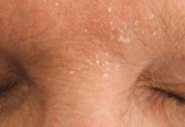
Seborrhoeic dermatitisSigns & symptomsInvestigationsDifferentialsTreatment algorithm | go to our full topic on Seborrhoeic dermatitis A common inflammatory skin disorder that usually manifests as erythema and scaling of the scalp, nasolabial folds, glabella, and occasionally central face and anterior chest. It tends to worsen with stress.[9] The adult scalp form is commonly termed dandruff or pityriasis capitis. Seborrhoeic dermatitis is common, with a prevalence of about 1% to 3% in adults living with immunocompetency.[10] It has a variable course that seldom completely subsides. An infant form (cradle cap) usually resolves within the first few months of life. Key risk factors include HIV infection, and aged <3 months. Characteristic history and physical examination findings are usually sufficient to diagnose the condition.[Figure caption and citation for the preceding image starts]: Seborrhoeic dermatitis, glabella, with scaling and mild erythemaPersonal collection of Dr Robert A. Schwartz [Citation ends]. |
Nappy rashSigns & symptomsInvestigationsDifferentialsTreatment algorithm | go to our full topic on Nappy rash Inflammation of the skin in the area of the body covered by a nappy. It is primarily an ICD. The key irritants are moisture from urine and faeces as well as faecal enzymes (ureases, proteases, and lipases). It is most common in the first 2 years of life, but can occur in any person who routinely wears nappies. Key risk factors include young age (<2 years), history of diarrhoea, underlying dermatological disorder, infrequent nappy changes, excess use of baby care products, plastic underpants, and no nappy-free time. Diagnosis is made by characteristic skin findings in the area of the body covered by a nappy; erythema of the convex surfaces of the buttocks is the classic finding. |
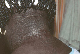
Lichen simplex chronicusSigns & symptomsInvestigationsDifferentialsTreatment algorithm | go to our full topic on Lichen simplex chronicus Lichen simplex chronicus (LCS; also known as neurodermatitis) is a common cutaneous disorder characterised by well-circumscribed erythematous, often hyperpigmented, patches and plaques of thickened lichenified skin. LSC is a common disease, with an estimated prevalence of 12%.[11] It most commonly occurs on the neck, ankles, scalp, pubis, vulva, scrotum, and extensor forearms as a result of chronic scratching and rubbing.[12] LSC patches or plaques can arise on skin affected by an underlying dermatosis such as atopic dermatitis, allergic contact dermatitis, stasis dermatitis, superficial fungal (tinea and candidiasis) and dermatophyte infections, lichen sclerosis, viral warts, scabies, lice, an arthropod bite, or a cutaneous neoplasia.[12][13] Several risk factors are associated with the development of LSC including atopic diathesis, environmental irritants, psychiatric disorders, and dermatological disease. Characteristic history and physical examination findings are normally sufficient for the diagnosis of LSC.[14] [Figure caption and citation for the preceding image starts]: Secondary lichen simplex chronicus in the setting of atopic dermatitisPersonal collection of Dr Swick [Citation ends]. |
SunburnSigns & symptomsInvestigationsDifferentialsTreatment algorithm | go to our full topic on Sunburn An acute inflammatory reaction of the skin induced by over-exposure to ultraviolet (UV) radiation that exceeds the individual’s minimal erythema dose. Skin findings include erythema and oedema, with or without vesiculation, followed by desquamation. Symptoms include pain and/or pruritus. In the UK, a 2022 survey found that 40% of adults reported at least one episode of sunburn in the past year, with this percentage increasing to 56% with young people (aged 18-34 years).[15] Key risk factors include Fitzpatrick skin types I-III, photo-sensitising drug, intentional tanning, and lack of or improper use of sunscreen. Primary prevention is critical, as cellular damage caused by UV radiation is irreversible and may with time increase the risk of skin cancer. Characteristic history and physical examination findings are usually sufficient to make the diagnosis of sunburn. |
Assessment of pruritusDifferentials | go to our full topic on Assessment of pruritus An unpleasant sensation that causes a desire to scratch. The most subjective symptom in dermatology is itching, which may occur with or without visible skin lesions. Pruritus can be either acute or chronic, with the chronic form lasting for 6 weeks or longer.[16] Chronic pruritus may be classified as being of dermatological, systemic, neurological, psychogenic/psychosomatic, mixed, or unknown aetiology. Pruritus is a subjective sensation; therefore, the diagnosis is based solely on the patient's symptoms. During clinical evaluation, it is important to identify a possible cause or disease responsible for itching, as well as determining the intensity and timeframe of the pruritus. A detailed history must also be collected regarding the concomitant symptoms, co-existent diseases, and medical problems, as well as drugs taken by the patient.[17] |
Assessment of rash in childrenDifferentials | go to our full topic on Assessment of rash in children Rash in children is common. A rash may be categorised as maculopapular, pustular, vesiculobullous, diffuse/erythematous, or petechial/purpuric in nature. Initial considerations in evaluating a rash in children include its morphology, duration, and distribution. Age, sex, family history, medications, known allergies, and exposures are also of primary importance. The primary differential diagnoses to consider for any rash presenting in childhood are viral exanthems, inflammatory dermatoses, local bacterial, fungal, or parasitic infections, tick-borne disease, drug eruptions, systemic bacterial infections, anaphylactic reactions, haematological disorders, and vasculitic and rheumatological conditions. |
Assessment of dermatological disorders in HIVDifferentials | go to our full topic on Assessment of dermatological disorders in HIV Dermatological disorders in HIV may be categorised as infectious, inflammatory, neoplastic, drug reaction, and metabolic. The dermatological manifestations of HIV are protean and often multiple in people living with HIV. HIV-specific dermatoses include HIV-related lipodystrophy, eosinophilic folliculitis, oral hairy leukoplakia, papular pruritic eruption of HIV, and HIV photodermatitis. Some skin diseases that appear in non-HIV-infected populations may have altered presentation in people living with HIV. Seborrhoeic dermatitis occurs with strikingly increased prevalence in people living with HIV.[18][19] Atopic dermatitis has a high prevalence in adult as well as paediatric populations with HIV.[20] Toxic epidermal necrolysis/Stevens-Johnson Syndrome must be recognised immediately, as this is a potentially life-threatening condition. |

Contributors
Authors
Editorial Team
BMJ Publishing Group
Disclosures
This overview has been compiled using the information in existing sub-topics.
References
Reference articles
A full list of sources referenced in this topic is available here.
Use of this content is subject to our disclaimer