Approach
Cryoglobulinaemia may be asymptomatic or present acutely. Mixed cryoglobulinaemia (MC) was first described as a triad of purpura, weakness, and arthralgia.[20] However, the clinical presentation of cryoglobulinaemia varies from asymptomatic to a cryoglobulinaemic syndrome consisting of purpura and leukocytoclastic vasculitis with multiple organ involvement.[4]
The pace and extent of evaluation depends on the presentation. Type l cryoglobulinaemia is usually associated with lymphoproliferative disorders. MC is associated with infectious or autoimmune disorders, particularly chronic hepatitis C virus (HCV) infection.[1][2] If a patient is diagnosed with a haematological malignancy while being evaluated for cryoglobulinaemia, referral to a haematologist is imperative.
Clinical evaluation
Careful clinical evaluation may provide clues to the type of cryoglobulinaemia and its complications:
The presence of acrocyanosis; Raynaud's phenomenon; skin ulcers; gangrene; and, less frequently, purpura, renal disease, or a neurological disorder should raise the possibility of type l cryoglobulinaemia.[1] This may be complicated by life-threatening hyperviscosity syndrome, which may present with bleeding, visual disturbances, diplopia, ataxia, headache, and confusion. Signs of retinal haemorrhage and retinal vein thrombosis may be seen.[21][22]
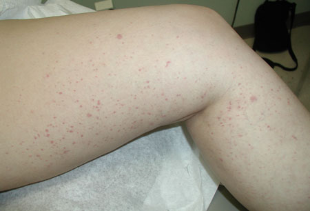
MC should be suspected in patients with signs and symptoms consistent with small- to medium-vessel vasculitis. These include the presence of purpura, mononeuritis multiplex, or glomerulonephritis.[23] It may suggest a more rapid work-up with particular attention to multiple organ function.[24] Other manifestations may include arthralgia, sicca syndrome, weakness, or hypertension in those with renal involvement.[Figure caption and citation for the preceding image starts]: Lower extremity palpable purpuraFrom the personal collection of Dr GS Kaeley [Citation ends].
 [Figure caption and citation for the preceding image starts]: Unusual case of hepatitis C-related cryoglobulin-induced digital gangreneAbdel-Gadir A, Patel, K, Dubrey SW. Cryoglobulinaemia induced digital gangrene in a case of hepatitis C. BMJ Case Reports. 2010 [Citation ends].
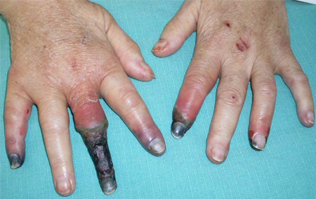
Isolation of cryoglobulins
The detection of serum cryoglobulins is necessary to classify the cryoglobulinaemic syndrome. Because cryoglobulins precipitate below normal body temperature, the collection and processing of the sample is critical.
Blood sampling (10-20 mL of blood is required), clotting, and serum separation should be carried out at 37°C (98.6°F). The serum sample is then stored at 4°C (39.2°F) for 24-72 hours and observed for the formation of a white precipitate (cryoglobulins). An aliquot of the cryoprecipitate should be re-warmed to 37°C (98.6°F) for 24 hours to test for the reversibility of cryoprecipitation.
Initial investigations
The Ig cryoprecipitate is isolated and analysed by immunoelectrophoresis or immunofixation. Cryoprecipitate measurement methods vary, and cryocrit levels do not correlate with disease activity.[1][4] However, a sudden decrease of cryoglobulin levels with increased levels of C4 may signal the evolution of a lymphoproliferative disorder.[25] This is thought to reflect the change of benign polyclonal B cells that produce antibodies to malignant B cells that no longer produce immunoglobulins.[4]
Other markers of activity are rheumatoid factor, complement C1q, C3, C4, CH50 (total complement activity), and acute-phase reactants (C-reactive protein and erythrocyte sedimentation rate [ESR]).[4][6][26] Findings may include IgM rheumatoid factor positive, elevated ESR, and normocytic normochromic anaemia. Complement C1q and C4 may be low.
The evaluation for organ involvement should include a full blood count, a comprehensive chemistry panel, and a urinalysis. An ECG and chest x-ray are indicated in patients with suspected multi-organ involvement.[4]
The aetiology of vasculitis should be assessed by performing the following tests: antinuclear antibodies (ANA), antineutrophil cytoplasmic antibodies, extractable nuclear antigen antibody (ENA), double-stranded deoxyribonucleic acid (dsDNA) antibodies, complement assay, and specific biopsies (e.g., IgA deposits on skin or kidney are suggestive of Henoch-Schonlein purpura).
If a viral cause is suspected, testing for HCV antibody and hepatitis B is recommended.[4]
Further investigations
Use of this content is subject to our disclaimer