Images and videos
Images


Assessment of tachycardia
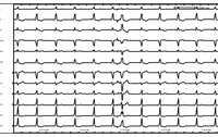
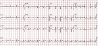
Atrial tachycardia: bursts of atrial tachycardia (10 beats in the middle section of the rhythm strip II at bottom) follows sinus complexes
From the collection of Dr Arti N. Shah
See this image in context in the following section/s:
Assessment of tachycardia
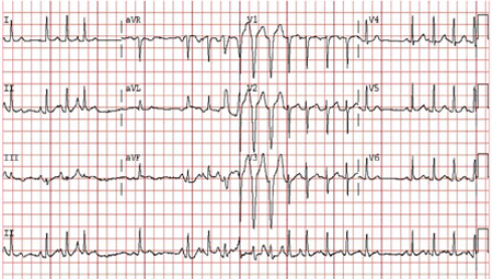
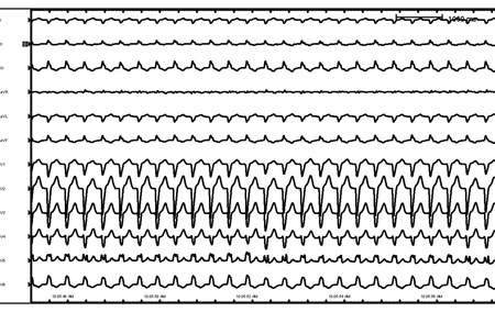
Right ventricular outflow tract ventricular tachycardia
From the collection of Robert W. Rho, MD; used with permission
See this image in context in the following section/s:
Assessment of tachycardia
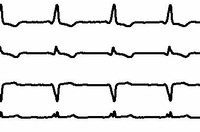
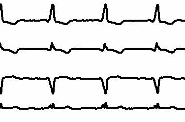
Atrial flutter (detail)
From the collection of Robert W. Rho, MD; used with permission
See this image in context in the following section/s:
Assessment of tachycardia
Sinus rhythm with pre-excitation
From the collection of Robert W. Rho, MD; used with permission
See this image in context in the following section/s:

Assessment of tachycardia
Antidromic re-entrant tachycardia
From the collection of Robert W. Rho, MD; used with permission
See this image in context in the following section/s:

Assessment of tachycardia
Atrial fibrillation: P waves are not discernible; the ventricular (QRS complexes) rate is irregularly irregular
From the collection of Dr Arti N. Shah
See this image in context in the following section/s:
Assessment of tachycardia
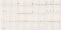
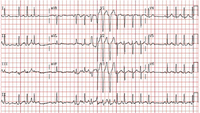
Multifocal atrial tachycardia
From the collections of Arti N. Shah and Bharat K. Kantharia
See this image in context in the following section/s:
Assessment of tachycardia
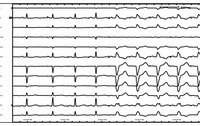
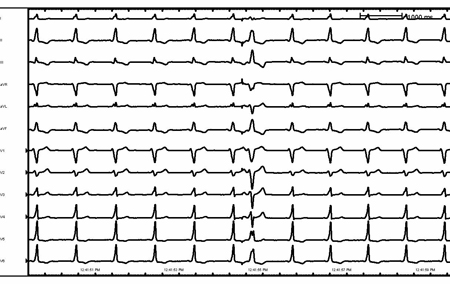
Supraventricular tachycardia with aberrancy and left bundle branch block
From the collection of Robert W. Rho, MD; used with permission
See this image in context in the following section/s:

Assessment of tachycardia
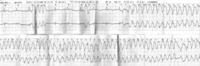
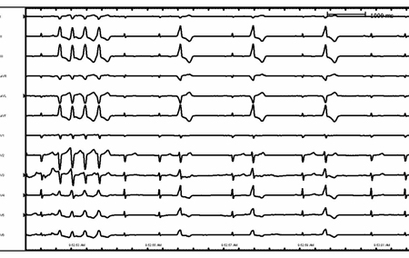
Ventricular tachycardia in a patient with arrhythmogenic right ventricular cardiomyopathy
From the collection of Robert W. Rho, MD; used with permission
See this image in context in the following section/s:

Assessment of tachycardia
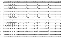
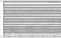
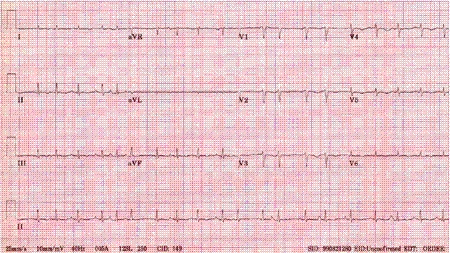
Artifact overlying sinus rhythm
From the collection of Robert W. Rho, MD; used with permission
See this image in context in the following section/s:
Assessment of tachycardia
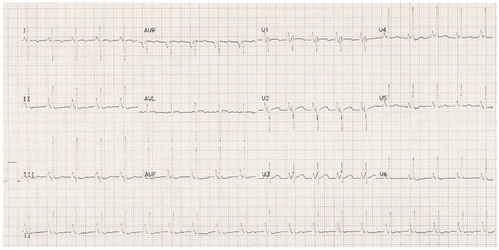
Atrial flutter: typical saw-tooth appearance of the flutter waves in the inferior leads (leads II, III, and aVF) indicates typical counterclockwise atrial flutter; the ventricular (QRS complexes) rate is variable
From the collection of Dr Arti N. Shah
See this image in context in the following section/s:
Assessment of tachycardia
Sinus rhythm with pre-excitation (detail)
From the collection of Robert W. Rho, MD; used with permission
See this image in context in the following section/s:

Assessment of tachycardia
Rate-related left bundle branch block
From the collection of Robert W. Rho, MD; used with permission
See this image in context in the following section/s:
Assessment of tachycardia
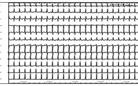
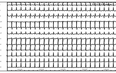
Atrial flutter
From the collection of Robert W. Rho, MD; used with permission
See this image in context in the following section/s:
Assessment of tachycardia
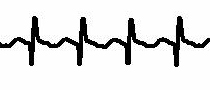
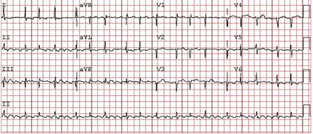
ECG example of sinus tachycardia
BMJ Learning/Professor Kevin Channer; used with permission
See this image in context in the following section/s:
Use of this content is subject to our disclaimer