Approach
Patients presenting with neurological dysfunction of the lower extremity require a careful history and exam to localise the lesion and identify the underlying cause. The character, localisation, duration, and severity of symptoms should be assessed, and clues to underlying systemic illness should be elucidated.
The aim of the exam is to identify the nerve(s) affected. Signs of systemic illness may be detected. Most compression neuropathies can be diagnosed clinically. If further investigation is needed, electrodiagnostic studies are usually the preferred investigation. Other investigations can be considered based on the clinical features.
Neurological history
It is important to characterise the neurological symptoms being experienced by the patient.[16]
Sensory symptoms: patients should be encouraged to describe their symptoms in detail in their own words. Common descriptions include 'boring', burning, stabbing, pins and needles, prickling, stinging, and sharp shooting pains. It is important to establish whether there is an associated loss of sensation, and whether it is in the same area as the paraesthesias.[17] Painful paraesthesias suggest an inflammatory or ischaemic process such as vasculitis. Shooting pains are characteristic of nerve entrapment.
Motor symptoms: associated symptoms of motor weakness or gait abnormalities (e.g., a foot drop) should also be assessed.
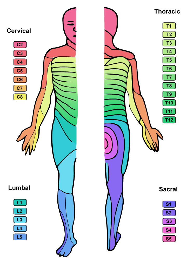
Localisation: the localisation of the symptoms provides clues as to the affected nerve and site of compression. Nerve root compression produces symptoms in the distribution of the affected dermatome and myotome. Plexopathies produce symptoms in the distribution of multiple peripheral nerves from the plexus. Compression or injury of isolated nerves distal to the plexus produces symptoms in the distribution of the individual nerve. [Figure caption and citation for the preceding image starts]: Dermatome mapAdapted by BMJ from an image by Ralf Stephan [Citation ends].
 [Figure caption and citation for the preceding image starts]: Cutaneous innervation of the leg, ankle and dorsum of footTeachMeAnatomy (https://teachmeanatomy.info/lower-limb/nerves/cutaneous-innervation/); used with permission [Citation ends].
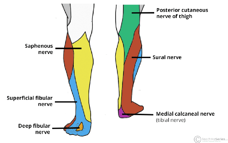
Duration and severity: the patient should be asked whether the symptoms are constant or relapsing and remitting, and whether any symptom has progressed. A history of similar previous symptoms should be sought.
When ruling out central nervous system (CNS) causes, the diagnosis of multiple sclerosis can be defined by the dissemination of lesions in distinct CNS anatomical locations and in multiple distinct clinical presentations.[18]
General medical history
Constitutional symptoms
Weight loss, night sweats, and/or fatigue may be present in infection, neoplastic disease, or a range of inflammatory conditions. Significant weight loss may lead to a compressive neuropathy.[19] Dry eyes or mouth may indicate Sjogren's syndrome.
Skin and joint changes
Ulcers, purpura, rash, or darkening of the skin may suggest peripheral vascular disease, infection, vasculitis, monoclonal protein production, or sarcoidosis. Arthralgias, joint swelling, or stiffness may indicate a rheumatological condition.
Past medical illnesses
It is important to establish whether the patient has any of the following underlying conditions: diabetes mellitus (level of control and complications of diabetic retinopathy and nephropathy), rheumatological disorders (SLE, Sjogren's syndrome, rheumatoid arthritis, vasculitides), cancer, or infectious disease (especially HIV).
If there is a history of diabetes mellitus or glucose-intolerant stages (e.g., pre-diabetes), diabetic amyotrophy should be suspected.[1] A history of serious antecedent illnesses or weight changes should be sought, although the patient may have had no additional symptoms. The patient may report pain in the pelvic/thigh region that is severe and 'boring'. The pain can sometimes resolve in 2 to 3 weeks before weakness and muscle atrophy are noted.
Establish any history of radiation therapy to the pelvis.
Family history
A positive family history of paraesthesias or sensory impairment may indicate an inherited neuropathy.
A family history of acquired diseases such as compression neuropathies, autoimmune disease, cancer, amyloidosis, or diabetes may also be present.
Social history
A history of smoking should raise suspicion of a paraneoplastic syndrome.
The presence of risk factors for HIV exposure or infection should prompt suspicion of HIV neuropathy.
Examination
Sensory system
The neurological examination should focus in detail on the areas that correspond to the patient's symptoms, but a general neurological examination should also be carried out to identify additional clues. The first step is to examine the sensory modalities in the lower extremities, with additional testing in the areas where the patient is reporting the paraesthesias.
Light touch may be tested with a cotton wisp. Temperature may be tested using a cold or warm stimulus. Pinprick may be tested using a single-use standard stimulus-producing tool such as a safety pin or similar sharp object. Joint position sense is tested using the distal interphalangeal joints of the great toe or middle finger. Vibration is tested using a 128 Hz tuning fork placed at the distal interphalangeal joint of the great toe or middle finger. The examination may reveal one of the following patterns.
Sensory loss in the distribution of a single peripheral nerve indicates a peripheral mononeuropathy. Palpation of an individual peripheral nerve or specific provocative testing, such as a Tinel’s sign, may elicit paraesthesias, indicating a nerve entrapment syndrome. Causes of compression, including malignancy, should be considered.
Symmetrical sensory loss in a glove-and-stocking distribution usually indicates a peripheral (length-dependent) polyneuropathy. This distribution is most common in diabetes mellitus and other metabolic disorders. It can also be seen in advanced cases of vasculitis.
Sensory loss in the distribution of multiple peripheral nerves indicates multiple mononeuropathies or a plexopathy. If it is painful, mononeuropathy multiplex should be considered and a cause for the vasculitis sought. Mononeuropathy multiplex may also indicate infection with HIV, cytomegalovirus (CMV), Herpes simplex virus (HSV), herpes zoster virus, Epstein-Barr virus (EBV), or leprosy. See Mononeuritis multiplex.
Sensory loss in a dermatomal pattern indicates a radiculopathy. The corresponding spinal nerve is also affected and the distribution of the spinal nerve should also be tested for sensory loss. If symptoms are painful, a radiculitis due to CMV, HSV, herpes zoster, or EBV may be present. Provocative testing may elicit symptoms but should not be relied on in isolation to establish the diagnosis.[20][21][22] [
 ]
]
Motor system
The presence of muscle atrophy should be noted. Weakness or atrophy in the same distribution as sensory loss indicates a mixed peripheral sensorimotor neuropathy (if the peripheral nerve distribution is affected) or involvement of a spinal root (if a dermatome and myotome are affected).
Loss of deep tendon reflexes may also be seen.
Gait should be assessed to determine whether there is a foot drop.
General examination
Fever may indicate infection.
Lymphadenopathy may indicate infection (e.g., HIV, HSV, or Lyme disease) or systemic inflammatory disease.
Diabetes mellitus should be considered if the patient is overweight or obese. Weight loss may be a sign of underlying malignancy.
Dry oral mucous membranes may indicate Sjogren's syndrome.
Purpura or rash suggests infection or vasculitis. Blistering in a dermatomal pattern indicates shingles. Changes in colour or thickening of the skin may indicate a monoclonal gammopathy or sarcoidosis.
Characteristic joint deformities may be present with underlying rheumatological conditions.
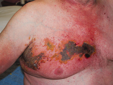
Funduscopy may reveal diabetic retinopathy in a diabetic patient.[Figure caption and citation for the preceding image starts]: Severe herpes zoster in an immunocompromised patient involving dermatomes T1 and T2BMJ Case Reports 2009 [doi:10.1136/bcr.07.2008.0533] Copyright © 2009 by the BMJ Publishing Group Ltd. [Citation ends].
 [Figure caption and citation for the preceding image starts]: Rheumatoid arthritis (chronic hand deformities)From the collection of Dr Soumya Chatterjee [Citation ends].
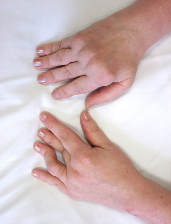
Initial testing
Electrodiagnostic studies
Electromyogram (EMG) and nerve conduction studies are frequently the first diagnostic tests ordered and should be directed at nerves and muscles whose involvement is suspected based on the clinical examination.[16]
EMG can characterise the type of peripheral nerve abnormality by its electrophysiological features (e.g., an axonal or demyelinating neuropathy). It can identify nerve entrapment syndromes and/or more proximal lesions such as radiculopathies or plexopathies and may be used to assess recovery.
Laboratory tests
If the clinical history, examination, or EMG and electrodiagnostic studies suggest the presence of systemic disease or more widespread nerve involvement (e.g., length-dependent polyneuropathy), initial lab tests such as FBC, serum urea nitrogen and creatinine, liver function testing, blood glucose testing with or without glucose tolerance testing, and thyroid function tests should be considered. If vasculitic, malignant, infectious, or inflammatory conditions are suspected, erythrocyte sedimentation rate, C-reactive protein, and complement levels should also be ordered.
Imaging
Neuromuscular ultrasound can confirm the location of the nerve lesion and identify structural causes of the focal neuropathy. Cross-sectional measurements of the nerve can be taken along the nerve, demonstrating an increase in cross-sectional area near (or just proximal to) the site of compression. As the lesion increases in severity (i.e., axon loss) the nerve may demonstrate loss of the normal fascicular architecture (axonotmesis) and even loss of continuity (neurotmesis). In severe axon loss lesions, muscles distal to the lesion may be more hyperechoic (light in colour) indicating muscle fibrosis. Structural causes of focal neuropathy that can be identified on ultrasound include an intraneural ganglion at the tibiofibular joint causing a common fibular neuropathy.[1][5][6]
If malignancy is suspected, a chest x-ray (particularly in smokers) may be ordered as part of the malignancy work-up, along with further imaging (i.e., CT scan of chest, abdomen, and pelvis) according to the suspected site and type of malignancy.
If a joint dislocation, trauma, or complications of surgery are suspected, plain films of the involved region should be ordered to exclude any orthopaedic emergencies.
Additional testing
Laboratory tests and pathology
Additional studies are ordered depending on the differential diagnosis after consideration of the history, examination, and electrodiagnostic testing and may include serological testing (e.g., HIV, HSV, EBV), lumbar puncture for cerebrospinal fluid (CSF) analysis, or biopsy (e.g., lymphoma).
Imaging
Neuro-imaging (MRI or CT) should be considered in any patient who is not responding to conservative treatment as expected, is displaying pain and dysfunction that appears out of proportion to the presumed mechanism of injury, or has signs or symptoms of systemic disease.
CT scan is useful for detecting retroperitoneal haemorrhages and evaluating bony structures.
MRI may be preferred for imaging soft tissues, compression sites, nerve roots, plexus, or individual named nerves.[23]
Interventional techniques
Referral to a spine interventionalist or pain management physician may be helpful in patients with suspected radiculopathy with negative or equivocal electrodiagnostic studies, and imaging. Guidelines by the American Society of Interventional Pain Physicians describe the evidence for the accuracy of lumbar spine diagnostic selective nerve blocks as ‘limited’; evidence for lumbar provocative discography is described as ‘fair’.[24]
Use of this content is subject to our disclaimer