Investigations
1st investigations to order
bilateral hip x-rays
Test
Anteroposterior and frog lateral views should be taken. Helps determine the stage of the disease process.[Figure caption and citation for the preceding image starts]: AP radiograph of a patient with Perthes' diseaseFrom the personal collection of Jwalant S. Mehta, MS (Orth), MCh (Orth), FRCS, FRCS (Orth) [Citation ends].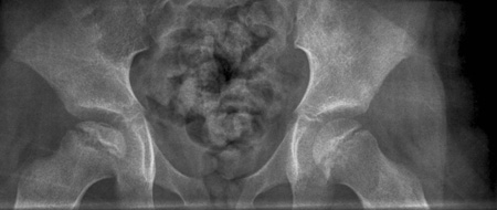 [Figure caption and citation for the preceding image starts]: Frog lateral radiograph of a patient with Perthes' diseaseFrom the personal collection of Jwalant S. Mehta, MS (Orth), MCh (Orth), FRCS, FRCS (Orth) [Citation ends].
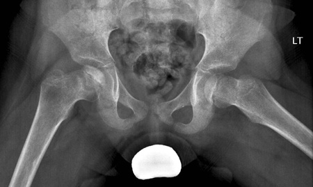
Result
femoral head collapse and fragmentation; subchondral fracture
Investigations to consider
FBC
Test
Consider in the acute phase to exclude other conditions.
Result
normal
serum erythrocyte sedimentation rate
Test
Consider in the acute phase to exclude other conditions.
Result
may be reactively increased in the symptomatic phase of the disease or may indicate an alternative pathology
serum C-reactive protein
Test
Consider in the acute phase to exclude other conditions.
Result
may be reactively increased in the symptomatic phase of the disease or may indicate an alternative pathology
bone scintigraphy
Test
Helps in the diagnosis during the ischaemic stage when radiographs may be normal.
Result
cold spot in the affected hip early in the disease process
MRI of hips
Test
Should be considered if radiographs appear normal. It is also a useful adjunct in the early stages of diagnosis, and has been found to be a sensitive modality in diagnosing Legg-Calvé-Perthes' disease.[58] If performed 6 months after disease onset, an MRI can accurately demonstrate the degree of epiphyseal involvement.[59] After re-ossification, MRI of the hips may also be useful in assessing the extent of the damage in one or both hips.[Figure caption and citation for the preceding image starts]: MRI scans of the same patient taken 8 years apart showing Perthes' disease of the left femoral epiphysis and proportional increase in cartilage thickness compared to the right side. Later scan (image on right) starting to show uptakeFrom the personal collection of Dominique Knight [Citation ends].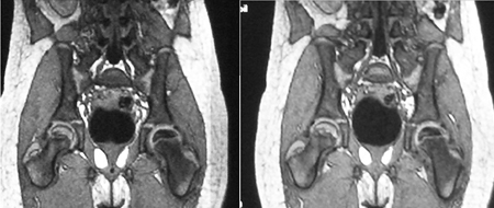 [Figure caption and citation for the preceding image starts]: MRI showing partial collapse of the left femoral head with areas of necrosisFrom BMJ Case Reports http://casereports.bmj.com/cgi/content/full/2009/jan08_1/bcr2007132811Copyright © 2011 by BMJ Publishing Group Ltd [Citation ends].
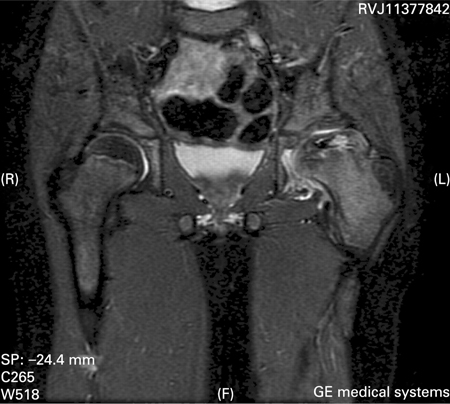
Result
femoral head collapse and fragmentation; prediction of final outcome with perfusion index
Use of this content is subject to our disclaimer