Images and videos
Images


Hirschsprung's disease
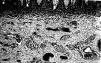
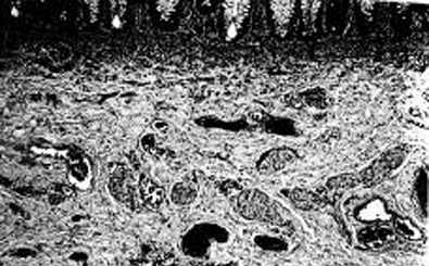
Histological section, including mucosa and submucosa of the rectum showing tortuous and hypertrophic nerve trunks of the submucosal plexus. There is no evidence of any ganglion cell present. This establishes the diagnosis of Hirschsprung's disease
Corman ML. Colon and rectal surgery. 5th ed. Philadelphia, PA: Lippincott Williams and Wilkins; 2005:555; used with permission
See this image in context in the following section/s:

Hirschsprung's disease
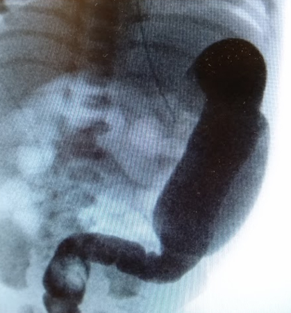
Barium enema performed in a newborn with Hirschsprung's disease. Often, classical changes are not obvious in the neonatal period
Corman ML. Colon and rectal surgery. 5th ed. Philadelphia, PA: Lippincott Williams and Wilkins; 2005:555-603; used with permission
See this image in context in the following section/s:

Hirschsprung's disease
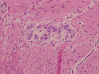
Hematoxylin and eosin showing ganglion cells in the myenteric plexus
From the personal collection of Lily Cheng, MD; used with permission
See this image in context in the following section/s:
Hirschsprung's disease
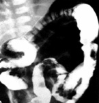
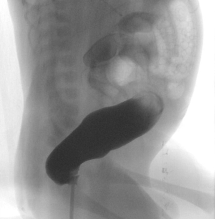
Contrast enema may demonstrate mucosal irregularity in the aganglionic distal colon and may show a transition zone between smaller caliber aganglionic distal colon and dilated proximal ganglionic colon
From the personal collection of Lily Cheng, MD; used with permission
See this image in context in the following section/s:
Hirschsprung's disease
Histological section, including mucosa with submucosa of the rectum showing clusters of ganglion cells in the submucosal plexus. This excludes Hirschsprung's disease at this level
Corman ML. Colon and rectal surgery. 5th ed. Philadelphia, PA: Lippincott Williams and Wilkins; 2005:555; used with permission
See this image in context in the following section/s:

Hirschsprung's disease
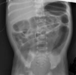
Abdominal x-ray image showing gaseous distension of the large bowel with air absent from the rectum (typical of Hirschsprung’s disease)
BMJ Case Reports 2012; doi:10.1136/bmj.e5521
See this image in context in the following section/s:
Hirschsprung's disease
Contrast enema showing an abnormal rectosigmoid ratio (sigmoid diameter larger than rectal diameter)
From the personal collection of Lily Cheng, MD; used with permission
See this image in context in the following section/s:
Hirschsprung's disease
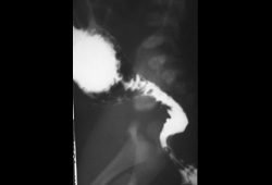
Contrast enema demonstrates the typical proximal dilation, transition zone, and non-distended, aganglionic portion
Corman ML. Colon and rectal surgery. 5th ed. Philadelphia, PA: Lippincott Williams and Wilkins; 2005:555-603; used with permission
See this image in context in the following section/s:
Use of this content is subject to our disclaimer