History and exam
Key diagnostic factors
common
presence of risk factors
Age <30 years, family history of vitiligo, and associated autoimmune diseases are strong risk factors for development of vitiligo. The disease most often associated with vitiligo is autoimmune thyroid disease.[31][30] Less commonly associated conditions include pernicious anaemia, SLE, alopecia areata, type 1 diabetes, and Addison's disease.
acral and periorificial depigmentation
Vitiligo is defined by round depigmented skin areas that have a predilection to acral and periorificial skin, but can also be confined to localised regions. [Figure caption and citation for the preceding image starts]: Typical periocular vitiligo with poliosis of several eyelashesPhotograph from the collection of Sarah Stein, MD, University of Chicago, used with permission [Citation ends].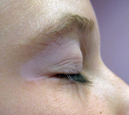
perianal and genital depigmentation
The presence of perianal and genital skin involvement may help to establish generalised disease. [Figure caption and citation for the preceding image starts]: Perianal and genital skin involvement in generalised vitiligoPhotograph from the collection of Sarah Stein, MD, University of Chicago, used with permission [Citation ends].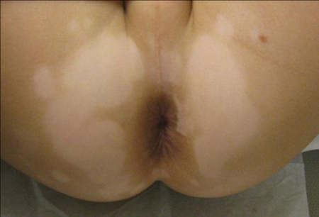
recent cutaneous trauma
Cutaneous trauma may be implicated in the appearance of new vitiligo or the spread of existing areas. Physical trauma (e.g., surgical incisions and friction) and sunburn have been widely described as triggers (Koebner's phenomenon).
localised sunburn pain
Depigmented skin is more susceptible to sunburn than unaffected areas.
enhancement and fluorescence with UV-A exposure
Wood's lamp accentuates the contrast between affected and unaffected skin (enhancement) while fluorescence is a characteristic bluish tint typical of vitiligo. [Figure caption and citation for the preceding image starts]: Accentuation of clinical features by Wood's lamp examinationFrom Dr Bernhard Ortel's personal collection [Citation ends].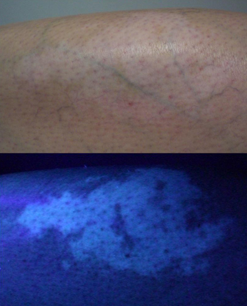
halo naevus
Halo (Sutton) naevus is found 10 times more commonly in patients with vitiligo.[31][Figure caption and citation for the preceding image starts]: Classic appearance of a halo naevusFrom Dr John E. Harris' personal collection [Citation ends].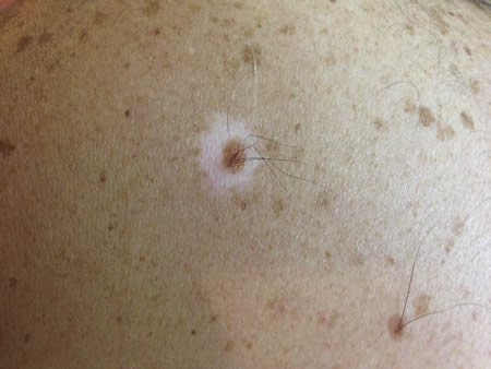 [Figure caption and citation for the preceding image starts]: Halo of depigmentation around congenital naevus in black childPhotograph from the collection of Sarah Stein, MD, University of Chicago, used with permission [Citation ends].
uncommon
universal depigmentation
Depigmentation of the vast majority of the body surface, so that the remaining unaffected skin appears as dark patches on a white skin background.
Risk factors
strong
age <30 years
family history of vitiligo
autoimmune disease
There is an increased incidence of autoimmune diseases in patients and relatives of vitiligo patients. Patients with such association may have an early onset of vitiligo.[31][30]
The disease most often associated with vitiligo is autoimmune thyroid disease.[31][30] Less commonly associated conditions include pernicious anaemia, systemic lupus erythematosus, alopecia areata, type 1 diabetes, and Addison's disease.
chemical contact
Exposure to a number of chemicals has been implicated in initiating or worsening vitiligo, including monobenzyl ether of hydroquinone, rhododendrol, and other phenols.[32]
One large population-based study implicated the use of permanent chemical hair dyes as risk factors for developing vitiligo, which supported earlier case reports.[33]
Use of this content is subject to our disclaimer