History and exam
Key diagnostic factors
common
presence of risk factors
Strong risk factors include prior occurrence of EM and herpetic or mycoplasma infection.
target lesions of the extremities
Lesions with 3 zones (red rim, clearance zone, and central blister or erosion) on the distal extremities are highly suggestive of EM.[2][3][4][5][Figure caption and citation for the preceding image starts]: Palmar target lesionsFrom the personal collection of Nanette Silverberg, MD; used with permission [Citation ends].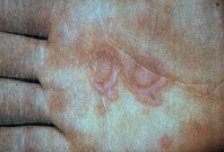
previous episode of EM
mucosal erosions
Seen in EM major. [Figure caption and citation for the preceding image starts]: Target lesions on the face and mucosal erosions with crusting due to HSV-1 recurrenceFrom the personal collection of Nanette Silverberg, MD; used with permission [Citation ends].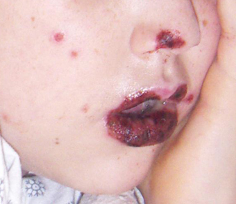
Other diagnostic factors
common
targetoid lesions

self-limiting course
clustered vesicles on an erythematous base
These may be seen if herpes simplex virus is the precipitating infection. [Figure caption and citation for the preceding image starts]: Target lesions on the face and mucosal erosions with crusting due to HSV-1 recurrenceFrom the personal collection of Nanette Silverberg, MD; used with permission [Citation ends].
rhonchi, rales, and/or wheezes
Lung findings are usually normal early in Mycoplasma pneumonia, but rhonchi, rales, and/or wheezes develop later on.
uncommon
red tympanic membranes
Strongly suggests Mycoplasma pneumonia if present.
Risk factors
strong
prior episode of erythema multiforme
While the exact mechanism of the hypersensitivity pattern of EM is unknown, recurrences are not uncommon. Thus, the strongest risk factor for occurrence is prior episodes of EM. In most cases, recurrence will relate to recurring herpes simplex virus infection.[36]
herpes simplex virus (HSV) infection
weak
certain other infections
Other infectious agents reported to trigger EM include cytomegalovirus, Epstein-Barr virus, SARS-CoV-2, hepatitis B virus, hepatitis C virus, influenza virus, HIV, herpes zoster, gardnerella, histoplasmosis (with concomitant erythema nodosum), coccidioidomycosis, orf (a disease of sheep and goats caused by a parapox virus that can be transmitted to humans), and syphilis.[2][7][10][11][12][13][14][15][16][37][38]
lymphoma
T-cell lymphoma may rarely mimic EM.[6] However, patients with lymphoma may have a dermatological manifestation of EM on presentation or during treatment.
Kawasaki disease
Case reports suggest an uncommon association between EM and Kawasaki disease.[17]
use of certain medicines
Drugs are reported as the cause of EM in less than 10% of cases, but a review of drug exposure is necessary for all patients.[2]
Drugs that have been associated with EM include certain antibiotics; docetaxel or paclitaxel; immune checkpoint inhibitors; sorafenib; tumour necrosis factor (TNF)-alpha inhibitors; antimalarials; hydroxychloroquine; lenalidomide; methotrexate; anticonvulsants; statins; bisphosphonates; non-steroidal anti-inflammatory drugs (NSAIDs); metamizole; oral contraceptives; imiquimod; lidocaine; triclocarban; barium contrast.[1][18][19][20][21][22][23][24][25][26][27] However, this list is not exhaustive and you should check your local drug information source.
Photo-distributed lesions have been noted with phenylbutazone, triclocarban, paclitaxel, and statins.[22][23]
One case report described the development of EM after 5 days of treatment with sorafenib; treatment was stopped and restarted 1 month later at a lower dose, but the patient experienced anaphylaxis 3 hours after ingesting the single dose of sorafenib.[27]
vaccinations
tattoos
Contact allergens have been known to trigger EM.[29]
Use of this content is subject to our disclaimer