Approach
Erythema nodosum (EN) predominantly affects women aged 20 to 40 years.[2] Diagnosis is clinical, but may be confirmed by biopsy, and should be accompanied by a search for the underlying cause.
Initial tests are performed to identify the most common and important causes, in particular streptococcal infections, sarcoidosis, and tuberculosis, plus any additional conditions prevalent in the geographical area. However, no cause is found in up to 60% of patients with erythema nodosum.
Skin lesions and physical examination
Lesions are typically red, tender, non-ulcerated, immobile nodules on the shins, typically resolving within 8 weeks of presentation. Though less common, lesions can also occur in areas other than the shins. Joint redness, swelling, and pain are often present.
In idiopathic cases, the chest, cardiovascular, abdominal, and other physical examinations are normal.
Patients with brucellosis or histoplasmosis may have an enlarged spleen and abnormal retinal examination. [Figure caption and citation for the preceding image starts]: Erythema nodosum on the shin of a patient.From the personal collection of Dr Om P. Sharma, MD, FRCP, DTM&H; used with permission [Citation ends].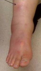 [Figure caption and citation for the preceding image starts]: Erythema nodosum on a patient's arms and handsCDC Public Health Image Library [Citation ends].
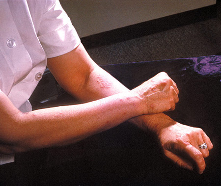 [Figure caption and citation for the preceding image starts]: Erythema nodosum lesions on skin of back due to hypersensitivity to antigens of Coccidioides immitisCDC Public Health Image Library [Citation ends].
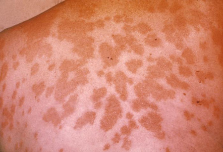 [Figure caption and citation for the preceding image starts]: Erythema nodosum on a patient's armFrom the collection of Dr Daniela Kroshinsky, MD, MPH; used with permission [Citation ends].
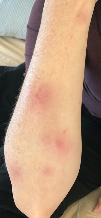 [Figure caption and citation for the preceding image starts]: Erythema nodosum on a patient's armFrom the collection of Dr Daniela Kroshinsky, MD, MPH; used with permission [Citation ends].
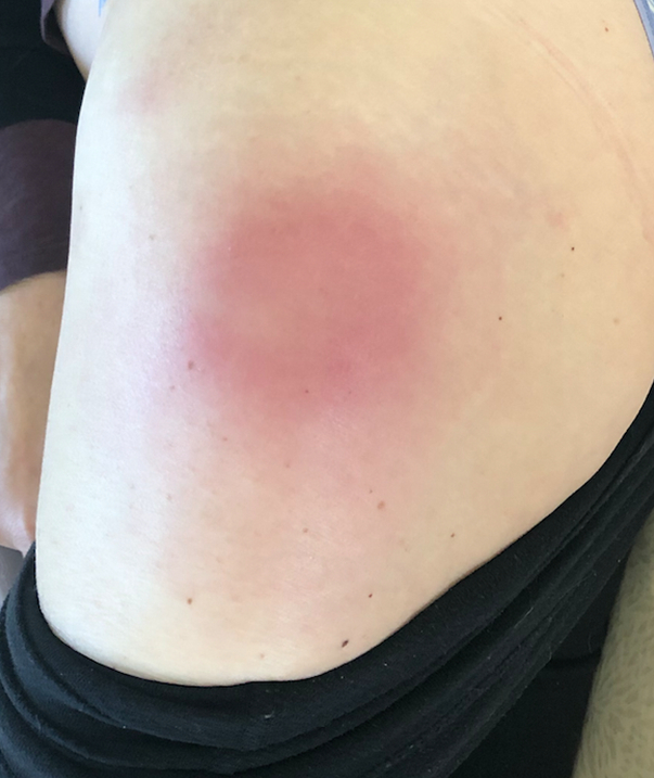
Initial investigations
FBC and anti-streptolysin-O (ASO) titres should be assessed on presentation and 2 to 4 weeks later. Chest x-ray (CXR) and tuberculin testing are also indicated initially in all patients.
Inflammation of the throat and tonsils may suggest streptococcal infection, even in the absence of elevated ASO titres.
Erythrocyte sedimentation rate is typically elevated in EN, but it is a non-specific marker of inflammation and so rarely affects the diagnosis or management.
Subsequent investigations
Subsequent testing is based on clinical suspicion of particular underlying conditions based on CXR and other test results, and various features of the history and physical examination:
CXR typically demonstrates bilateral hilar adenopathy in sarcoidosis and unilateral hilar node enlargement in tuberculosis, coccidioidomycosis, and brucellosis. In psittacosis pneumonia, CXR shows pneumonic infiltrate in the lower lobes.
For sarcoidosis, diagnosis may be made clinically. Lofgren's syndrome is the presentation of uveitis, arthralgias, and fever with EN nodules. Serum ACE is elevated in about 60% of sarcoidosis patients.
If tuberculin skin test or interferon gamma release assay is negative, and tuberculosis is no longer suspected, unilateral hilar node enlargement on CXR may prompt further investigation of a number of possible aetiologies, depending on additional historical, geographical, and physical examination features. Coccidioidin or histoplasmin skin tests, serological testing for blastomycosis, and serological testing for brucellosis or psittacosis, may be indicated.
Concomitant gastrointestinal symptoms raise suspicion of underlying inflammatory bowel disease, Behcet's disease, or Yersinia infection. Inflammatory bowel disease may be investigated with endoscopy. Serum IgD level is high in Behcet's disease. Serum agglutinins and stool cultures are positive in Yersinia infections.
Presence of joint symptoms or signs may suggest rheumatological disease as the underlying cause. This may be further investigated by taking x-rays of affected joints, rheumatoid factor testing, or other testing as indicated by the clinical picture.
Cancer is a rare aetiology. The patient’s history and physical examination findings should be used to help guide further testing for possible underlying malignancy.
Skin biopsy
EN is primarily a clinical diagnosis, but biopsy may be indicated for atypical lesions, such as those that demonstrate purpura or ulceration, have an atypical distribution, or in unresponsive cases. In such instances, biopsy may help distinguish EN from other conditions characterised by panniculitis, such as lupus panniculitis, pancreatic fat necrosis, nodular vasculitis, Weber-Christian disease, superficial thrombophlebitis, factitial disease, alpha-1 anti-trypsin deficiency, lymphoma, and infection.[2]
Of note, it is important to have an adequate sample of the subcutaneous fat. This can be accomplished in most cases with a punch biopsy; however, a small incisional biopsy may be needed if results are indeterminate.[Figure caption and citation for the preceding image starts]: Pancreatic panniculitis in a 56-year-old male with neurofibromatosis 2 (NF2) and erythema nodosum (prednisolone 10 mg daily)From the collection of Dr Daniela Kroshinsky, MD, MPH; used with permission [Citation ends].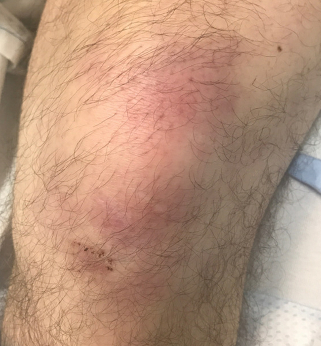 [Figure caption and citation for the preceding image starts]: Pancreatic panniculitis in a 56-year-old male with neurofibromatosis 2 (NF2) and erythema nodosum (prednisolone 10 mg daily)From the collection of Dr Daniela Kroshinsky, MD, MPH; used with permission [Citation ends].
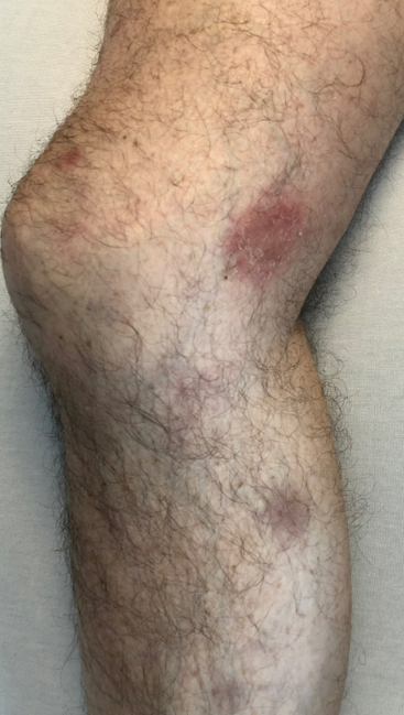 [Figure caption and citation for the preceding image starts]: Subcutaneous panniculitis-like T cell lymphoma in a 69-year-old female with hepatitis B, thyroid cancer, and recent insect exposure while gardeningFrom the collection of Dr Daniela Kroshinsky, MD, MPH; used with permission [Citation ends].
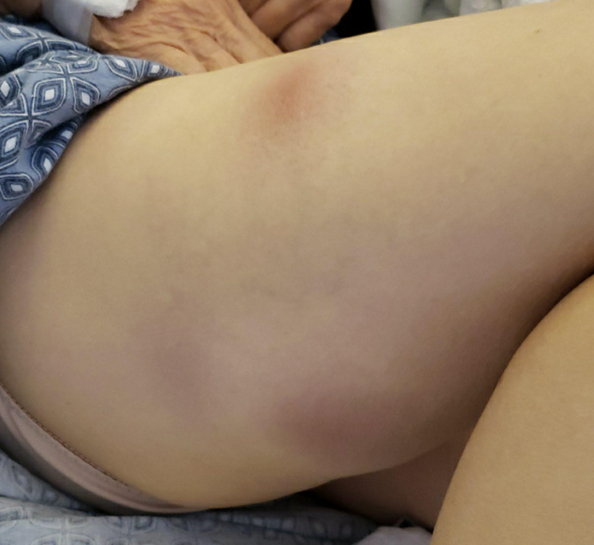 [Figure caption and citation for the preceding image starts]: Subacute nodular migratory panniculitisFrom the collection of Dr Daniela Kroshinsky, MD, MPH; used with permission [Citation ends].
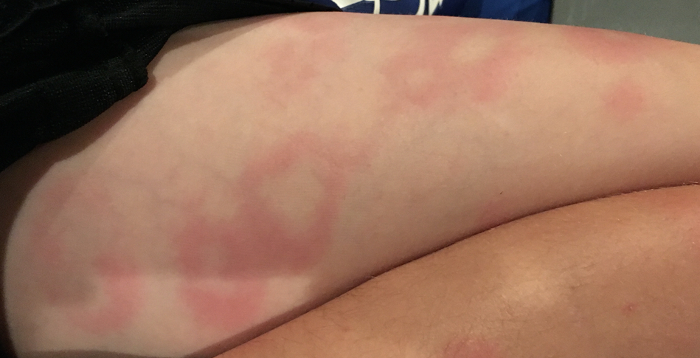
Use of this content is subject to our disclaimer