Non-Hodgkin's lymphomas (NHLs) are a heterogeneous group of malignancies with over 60 subtypes.[1]Alaggio R, Amador C, Anagnostopoulos I, et al. The 5th edition of the World Health Organization classification of haematolymphoid tumours: lymphoid neoplasms. Leukemia. 2022 Jul;36(7):1720-48.
https://www.nature.com/articles/s41375-022-01620-2
http://www.ncbi.nlm.nih.gov/pubmed/35732829?tool=bestpractice.com
[2]Campo E, Jaffe ES, Cook JR, et al. The International Consensus Classification of mature lymphoid neoplasms: a report from the Clinical Advisory Committee. Blood. 2022 Sep 15;140(11):1229-53.
https://pmc.ncbi.nlm.nih.gov/articles/PMC9479027
http://www.ncbi.nlm.nih.gov/pubmed/35653592?tool=bestpractice.com
See Classification.
Patients with NHL may present differently depending on the type and location of lymphoma, and disease stage at presentation.
Diagnosis of NHL is based on history, physical examination, laboratory tests, tissue biopsy, immunophenotyping (immunohistochemistry and/or flow cytometry), and imaging (e.g., positron emission tomography/computed tomography [PET/CT]).[70]National Comprehensive Cancer Network. NCCN clinical practice guidelines in oncology: B-cell lymphomas [internet publication].
https://www.nccn.org/guidelines/category_1
[71]National Comprehensive Cancer Network. NCCN clinical practice guidelines in oncology: T-cell lymphomas [internet publication].
https://www.nccn.org/guidelines/category_1
Genetic studies should be considered to identify genetic abnormalities that can guide diagnosis, prognosis, and treatment.[70]National Comprehensive Cancer Network. NCCN clinical practice guidelines in oncology: B-cell lymphomas [internet publication].
https://www.nccn.org/guidelines/category_1
[71]National Comprehensive Cancer Network. NCCN clinical practice guidelines in oncology: T-cell lymphomas [internet publication].
https://www.nccn.org/guidelines/category_1
NHL may mimic many other conditions and can be difficult to distinguish from inflammation, benign hyperplasia, carcinomas, germ cell tumours, or melanoma.
Given the complexity and heterogeneity of NHL, an expert haematopathologist should be consulted to establish an accurate diagnosis in patients with clinical features that do not fit the pathological diagnosis, or in the setting of difficult histological diagnoses. Examples may include patients with:
Features of both primary mediastinal B-cell lymphoma and classic Hodgkin's lymphoma (i.e., mediastinal grey zone lymphoma)
Features of both diffuse large B-cell lymphoma (DLBCL) and Burkitt's lymphoma
Suspected primary cutaneous DLBCL, leg type
T-cell lymphomas
Composite lymphomas (concurrent involvement of two types of lymphomas)
or where there is a need to differentiate:
History and physical examination
History and findings on physical examination will vary depending on the type and location of lymphoma, and disease stage at presentation.
History may reveal risk factors associated with NHL, such as:
Prior viral infection (e.g., HIV, Epstein-Barr virus [EBV], hepatitis C virus [HCV], human T-cell lymphotrophic virus type 1 [HTLV-1])
Bacterial infection (e.g., Coxiella burnetii)
Autoimmune disorder (e.g., Sjogren syndrome, rheumatoid arthritis)
Immunodeficiency due to a disorder (e.g., common variable immunodeficiency) or acquired (e.g., post-transplant)
Genetic disorder (e.g., Klinefelter syndrome)
Exposure to certain chemicals (e.g., pesticides)
Use of immunomodulatory drugs, such as tumour necrosis factor (TNF)-alpha antagonists (e.g., infliximab, adalimumab)
Breast implant (particularly if textured)
Clinical presentation
Clinical presentation is often vague and can be very diverse, ranging from asymptomatic or minimally symptomatic (e.g., painless enlarged lymph nodes) in those with an indolent NHL (e.g., follicular lymphoma), to acute presentation in those with an aggressive NHL (e.g., DLBCL).
Patients with aggressive (high-grade) NHL or advanced-stage disease may present with the following:[3]Hingorjo MR, Syed S. Presentation, staging and diagnosis of lymphoma: a clinical perspective. J Ayub Med Coll Abbottabad. 2008 Oct-Dec;20(4):100-3.
http://www.ncbi.nlm.nih.gov/pubmed/19999217?tool=bestpractice.com
[4]Wang G, Chang Y, Wu X, et al. Clinical features and prognostic factors of primary bone marrow lymphoma. Cancer Manag Res. 2019;11:2553-63.
https://pmc.ncbi.nlm.nih.gov/articles/PMC6446986
http://www.ncbi.nlm.nih.gov/pubmed/31015766?tool=bestpractice.com
[5]Yu Y, Dong X, Tu M, et al. Primary mediastinal large B cell lymphoma. Thorac Cancer. 2021 Nov;12(21):2831-7.
https://pmc.ncbi.nlm.nih.gov/articles/PMC8563158
http://www.ncbi.nlm.nih.gov/pubmed/34590432?tool=bestpractice.com
[6]Savage KJ, Chhanabhai M, Gascoyne RD, et al. Characterization of peripheral T-cell lymphomas in a single North American institution by the WHO classification. Ann Oncol. 2004 Oct;15(10):1467-75.
https://www.annalsofoncology.org/article/S0923-7534(19)51047-X/fulltext
http://www.ncbi.nlm.nih.gov/pubmed/15367405?tool=bestpractice.com
[7]Cadranel J, Wislez M, Antoine M. Primary pulmonary lymphoma. Eur Respir J. 2002 Sep;20(3):750-62.
https://publications.ersnet.org/content/erj/20/3/750
http://www.ncbi.nlm.nih.gov/pubmed/12358356?tool=bestpractice.com
[8]Kaji D, Ota Y, Sato Y, et al. Primary human herpesvirus 8-negative effusion-based lymphoma: a large B-cell lymphoma with favorable prognosis. Blood Adv. 2020 Sep 22;4(18):4442-50.
https://pmc.ncbi.nlm.nih.gov/articles/PMC7509864
http://www.ncbi.nlm.nih.gov/pubmed/32936906?tool=bestpractice.com
[9]Khdhir M, El Annan T, El Amine MA, et al. Complications of lymphoma in the abdomen and pelvis: clinical and imaging review. Abdom Radiol (NY). 2022 Aug;47(8):2937-55.
https://pmc.ncbi.nlm.nih.gov/articles/PMC10509750
http://www.ncbi.nlm.nih.gov/pubmed/35690955?tool=bestpractice.com
[10]Schaff LR, Grommes C. Primary central nervous system lymphoma. Blood. 2022 Sep 1;140(9):971-9.
https://pmc.ncbi.nlm.nih.gov/articles/PMC7266949
http://www.ncbi.nlm.nih.gov/pubmed/34699590?tool=bestpractice.com
[11]Müller A, Dreyling M, Roeder F, et al. Primary bone lymphoma: clinical presentation and therapeutic considerations. J Bone Oncol. 2020 Dec;25:100326.
https://pmc.ncbi.nlm.nih.gov/articles/PMC7554647
http://www.ncbi.nlm.nih.gov/pubmed/33083218?tool=bestpractice.com
[12]Monnard V, Sun A, Epelbaum R, et al. Primary spinal epidural lymphoma: patients' profile, outcome, and prognostic factors: a multicenter Rare Cancer Network study. Int J Radiat Oncol Biol Phys. 2006 Jul 1;65(3):817-23.
http://www.ncbi.nlm.nih.gov/pubmed/16542791?tool=bestpractice.com
[13]Jaffe ES, Ashar BS, Clemens MW, et al. Best practices guideline for the pathologic diagnosis of breast implant-associated anaplastic large-cell lymphoma. J Clin Oncol. 2020 Apr 1;38(10):1102-11.
https://ascopubs.org/doi/10.1200/JCO.19.02778
http://www.ncbi.nlm.nih.gov/pubmed/32045544?tool=bestpractice.com
[14]Opyrchal M, Figanbaum T, Ghosh A, et al. Spontaneous tumor lysis syndrome in the setting of B-cell lymphoma. Case Rep Med. 2010;2010:610969.
https://pmc.ncbi.nlm.nih.gov/articles/PMC2836528
http://www.ncbi.nlm.nih.gov/pubmed/20300188?tool=bestpractice.com
[15]Belay Y, Yirdaw K, Enawgaw B. Tumor lysis syndrome in patients with hematological malignancies. J Oncol. 2017;2017:9684909.
https://pmc.ncbi.nlm.nih.gov/articles/PMC5688348
http://www.ncbi.nlm.nih.gov/pubmed/29230244?tool=bestpractice.com
B symptoms (unexplained fever, drenching night sweats, and weight loss >10% of body weight within 6 months)
Fatigue/malaise
Chest pain
Shortness of breath and/or pallor (due to anaemia; pulmonary or pleural involvement; pleural/pericardial effusion)
Cough (due to pulmonary, mediastinal, or lymph node involvement)
Abdominal discomfort or pain (due to gastrointestinal, liver, spleen, or lymph node involvement)
Headache/change in mental status (due to meningeal or parenchymal brain involvement)
Focal neurological deficits; for example, ataxia, cognitive changes, and focal weakness (due to central nervous system [CNS] involvement)
Bone pain (due to bone involvement)
Back pain (due to spinal bone or epidural involvement)
Breast pain (due to breast implant involvement)
Metabolic abnormalities (acute renal injury, tumour lysis syndrome, hypercalcaemia)
Physical examination may identify the following:[3]Hingorjo MR, Syed S. Presentation, staging and diagnosis of lymphoma: a clinical perspective. J Ayub Med Coll Abbottabad. 2008 Oct-Dec;20(4):100-3.
http://www.ncbi.nlm.nih.gov/pubmed/19999217?tool=bestpractice.com
[10]Schaff LR, Grommes C. Primary central nervous system lymphoma. Blood. 2022 Sep 1;140(9):971-9.
https://pmc.ncbi.nlm.nih.gov/articles/PMC7266949
http://www.ncbi.nlm.nih.gov/pubmed/34699590?tool=bestpractice.com
[13]Jaffe ES, Ashar BS, Clemens MW, et al. Best practices guideline for the pathologic diagnosis of breast implant-associated anaplastic large-cell lymphoma. J Clin Oncol. 2020 Apr 1;38(10):1102-11.
https://ascopubs.org/doi/10.1200/JCO.19.02778
http://www.ncbi.nlm.nih.gov/pubmed/32045544?tool=bestpractice.com
[72]Franco V, Florena AM, Iannitto E. Splenic marginal zone lymphoma. Blood. 2003 Apr 1;101(7):2464-72.
https://www.sciencedirect.com/science/article/pii/S0006497120511269?via%3Dihub
http://www.ncbi.nlm.nih.gov/pubmed/12446449?tool=bestpractice.com
[73]Vitiello P, Sica A, Ronchi A, et al. Primary cutaneous B-cell lymphomas: an update. Front Oncol. 2020;10:651.
https://pmc.ncbi.nlm.nih.gov/articles/PMC7266949
http://www.ncbi.nlm.nih.gov/pubmed/32528871?tool=bestpractice.com
Laboratory tests
Routine laboratory tests include:[70]National Comprehensive Cancer Network. NCCN clinical practice guidelines in oncology: B-cell lymphomas [internet publication].
https://www.nccn.org/guidelines/category_1
[71]National Comprehensive Cancer Network. NCCN clinical practice guidelines in oncology: T-cell lymphomas [internet publication].
https://www.nccn.org/guidelines/category_1
[74]Forkasiewicz A, Dorociak M, Stach K, et al. The usefulness of lactate dehydrogenase measurements in current oncological practice. Cell Mol Biol Lett. 2020;25:35.
https://www.doi.org/10.1186/s11658-020-00228-7
http://www.ncbi.nlm.nih.gov/pubmed/32528540?tool=bestpractice.com
[75]Fox CP, Chaganti S, McIlroy G, et al. The management of newly diagnosed large B-cell lymphoma: a British Society for Haematology Guideline. Br J Haematol. 2024 Apr;204(4):1178-92.
https://pmc.ncbi.nlm.nih.gov/articles/PMC7616447
http://www.ncbi.nlm.nih.gov/pubmed/38247115?tool=bestpractice.com
Full blood count (FBC) with differential
Comprehensive metabolic panel (including liver function tests [LFTs])
Serum lactate dehydrogenase (LDH), an indirect measure of the proliferative rate of the lymphoma
Uric acid (particularly for aggressive lymphomas)
The above laboratory tests are required for the purpose of:
Assessing blood and organ function (e.g., liver, kidney)
Guiding diagnosis and treatment (including tumour lysis syndrome prophylaxis)
Risk assessment and prognostication (see Criteria)
Monitoring disease course
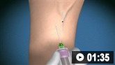
Biopsy
Excisional or incisional lymph node biopsy should be performed for diagnosis and grading of NHL, as this allows for optimal assessment of morphology (including lymph node architecture) and provides sufficient tissue for immunohistochemistry and molecular studies.[70]National Comprehensive Cancer Network. NCCN clinical practice guidelines in oncology: B-cell lymphomas [internet publication].
https://www.nccn.org/guidelines/category_1
[71]National Comprehensive Cancer Network. NCCN clinical practice guidelines in oncology: T-cell lymphomas [internet publication].
https://www.nccn.org/guidelines/category_1
[77]Kroft SH, Sever CE, Bagg A, et al. Laboratory workup of lymphoma in adults: guideline from the American Society for Clinical Pathology and the College of American Pathologists. Arch Pathol Lab Med. 2021 Mar 1;145(3):269-90.
https://meridian.allenpress.com/aplm/article/145/3/269/447620/Laboratory-Workup-of-Lymphoma-in-Adults-Guideline
http://www.ncbi.nlm.nih.gov/pubmed/33175094?tool=bestpractice.com
Core needle biopsy is an appropriate alternative to excisional or incisional biopsy in certain circumstances (e.g., if a lymph node is not easily accessible, or if surgery is not possible or will substantially delay treatment).[70]National Comprehensive Cancer Network. NCCN clinical practice guidelines in oncology: B-cell lymphomas [internet publication].
https://www.nccn.org/guidelines/category_1
[71]National Comprehensive Cancer Network. NCCN clinical practice guidelines in oncology: T-cell lymphomas [internet publication].
https://www.nccn.org/guidelines/category_1
FNA biopsy alone is insufficient for diagnosis and grading of NHL.[70]National Comprehensive Cancer Network. NCCN clinical practice guidelines in oncology: B-cell lymphomas [internet publication].
https://www.nccn.org/guidelines/category_1
[71]National Comprehensive Cancer Network. NCCN clinical practice guidelines in oncology: T-cell lymphomas [internet publication].
https://www.nccn.org/guidelines/category_1
It may be used for the initial diagnostic work-up for certain lymphomas (e.g., breast implant-associated anaplastic large cell lymphoma [BIA-ALCL]).[13]Jaffe ES, Ashar BS, Clemens MW, et al. Best practices guideline for the pathologic diagnosis of breast implant-associated anaplastic large-cell lymphoma. J Clin Oncol. 2020 Apr 1;38(10):1102-11.
https://ascopubs.org/doi/10.1200/JCO.19.02778
http://www.ncbi.nlm.nih.gov/pubmed/32045544?tool=bestpractice.com
[71]National Comprehensive Cancer Network. NCCN clinical practice guidelines in oncology: T-cell lymphomas [internet publication].
https://www.nccn.org/guidelines/category_1
[78]Turton P, El-Sharkawi D, Lyburn I, et al. UK guidelines on the diagnosis and treatment of breast implant-associated anaplastic large cell lymphoma on behalf of the Medicines and Healthcare products Regulatory Agency Plastic, Reconstructive and Aesthetic Surgery Expert Advisory Group. Br J Haematol. 2021 Feb;192(3):444-58.
https://pmc.ncbi.nlm.nih.gov/articles/PMC7894347
http://www.ncbi.nlm.nih.gov/pubmed/33222158?tool=bestpractice.com
Biopsy of extranodal sites
Biopsy of extranodal sites may be required for diagnosising certain types of NHL, for example:[71]National Comprehensive Cancer Network. NCCN clinical practice guidelines in oncology: T-cell lymphomas [internet publication].
https://www.nccn.org/guidelines/category_1
[79]National Comprehensive Cancer Network. NCCN central nervous system cancers [internet publication].
https://www.nccn.org/guidelines/category_1
[80]National Comprehensive Cancer Network. NCCN clinical practice guidelines in oncology: primary cutaneous lymphomas [internet publication].
https://www.nccn.org/guidelines/category_1
Stereotactic biopsy of brain lesions for diagnosing primary CNS lymphoma
FNA biopsy of periprosthetic effusion (>50 mL) and surgical biopsy (excisional, incisional, or core needle) of a breast mass for diagnosing BIA-ALCL
Skin biopsy (punch, incisional, or excisional) for diagnosing primary cutaneous lymphomas, or in cases of skin involvement by other lymphomas
Vitreous fluid biopsy for primary vitreoretinal lymphoma or primary CNS lymphoma ocular variant
Bone marrow biopsy and aspiration may be helpful for evaluating unexplained cytopenias and establishing a diagnosis (including staging), depending on the type of NHL or the specific setting (e.g., when lymph node biopsy is not diagnostic and bone marrow involvement is suspected).[70]National Comprehensive Cancer Network. NCCN clinical practice guidelines in oncology: B-cell lymphomas [internet publication].
https://www.nccn.org/guidelines/category_1
[77]Kroft SH, Sever CE, Bagg A, et al. Laboratory workup of lymphoma in adults: guideline from the American Society for Clinical Pathology and the College of American Pathologists. Arch Pathol Lab Med. 2021 Mar 1;145(3):269-90.
https://meridian.allenpress.com/aplm/article/145/3/269/447620/Laboratory-Workup-of-Lymphoma-in-Adults-Guideline
http://www.ncbi.nlm.nih.gov/pubmed/33175094?tool=bestpractice.com
Assessing bone marrow involvement is important for staging and guiding treatment (e.g., stem cell transplant).[81]Cheson BD, Fisher RI, Barrington SF, et al. Recommendations for initial evaluation, staging, and response assessment of Hodgkin and non-Hodgkin lymphoma: the Lugano classification. J Clin Oncol. 2014 Sep 20;32(27):3059-68.
https://pmc.ncbi.nlm.nih.gov/articles/PMC4979083
http://www.ncbi.nlm.nih.gov/pubmed/25113753?tool=bestpractice.com
Bone marrow involvement usually signifies stage IV disease. See Criteria.
Immunophenotyping
Immunohistochemistry and/or flow cytometry of the biopsy specimen should be performed to identify tumour markers to confirm the type of NHL.[70]National Comprehensive Cancer Network. NCCN clinical practice guidelines in oncology: B-cell lymphomas [internet publication].
https://www.nccn.org/guidelines/category_1
[71]National Comprehensive Cancer Network. NCCN clinical practice guidelines in oncology: T-cell lymphomas [internet publication].
https://www.nccn.org/guidelines/category_1
Flow cytometry is particularly useful when tumour cells are suspended (e.g., in peripheral blood, bone marrow aspirate, lymph node suspensions, effusions, cerebrospinal fluid)
Genetic studies
The following genetic studies can be used to detect genetic abnormalities associated with NHL, which can help establish a diagnosis (e.g., by confirming a malignant clone and determining the NHL subtype) and guide prognosis and treatment:[70]National Comprehensive Cancer Network. NCCN clinical practice guidelines in oncology: B-cell lymphomas [internet publication].
https://www.nccn.org/guidelines/category_1
[71]National Comprehensive Cancer Network. NCCN clinical practice guidelines in oncology: T-cell lymphomas [internet publication].
https://www.nccn.org/guidelines/category_1
Molecular analysis (e.g., polymerase chain reaction [PCR]) to detect immunoglobulin gene rearrangements (in B-cell lymphomas) or T-cell receptor gene rearrangements (in T-cell lymphomas)
Cytogenetic analysis (e.g., karyotype; fluorescence in situ hybridisation [FISH]) to detect chromosome translocations/rearrangements involving oncogenes such as BCL2 (e.g., t(14;18) in follicular lymphoma and DLBCL), CCND1 (e.g., t(11;14) in mantle cell lymphoma), MYC (e.g., t(8;14) in Burkitt's lymphoma and DLBCL), and BCL6 (e.g., t(3;14) in DLBCL)
Mutational analysis (e.g., gene sequencing; next-generation sequencing [NGS]) to detect genetic mutations (e.g., TP53 in mantle cell lymphoma)
Imaging
Fluorodeoxyglucose (FDG)-PET/CT scan or CT scan alone (of chest, abdomen, pelvis, head/neck [in some cases]) should be performed as part of the standard work-up (diagnosis, staging) and follow-up of NHL.[70]National Comprehensive Cancer Network. NCCN clinical practice guidelines in oncology: B-cell lymphomas [internet publication].
https://www.nccn.org/guidelines/category_1
[71]National Comprehensive Cancer Network. NCCN clinical practice guidelines in oncology: T-cell lymphomas [internet publication].
https://www.nccn.org/guidelines/category_1
[79]National Comprehensive Cancer Network. NCCN central nervous system cancers [internet publication].
https://www.nccn.org/guidelines/category_1
[80]National Comprehensive Cancer Network. NCCN clinical practice guidelines in oncology: primary cutaneous lymphomas [internet publication].
https://www.nccn.org/guidelines/category_1
FDG-PET/CT scan is the preferred imaging modality for staging and end-of-treatment response assessment in patients with FDG-avid lymphomas (e.g., DLBCL, follicular lymphoma) as it is more accurate than CT scan alone in detecting nodal and extranodal lesions.[70]National Comprehensive Cancer Network. NCCN clinical practice guidelines in oncology: B-cell lymphomas [internet publication].
https://www.nccn.org/guidelines/category_1
[71]National Comprehensive Cancer Network. NCCN clinical practice guidelines in oncology: T-cell lymphomas [internet publication].
https://www.nccn.org/guidelines/category_1
[82]Schaefer NG, Hany TF, Taverna C, et al. Non-Hodgkin lymphoma and Hodgkin disease: coregistered FDG PET and CT at staging and restaging--do we need contrast-enhanced CT? Radiology. 2004 Sep;232(3):823-9.
http://www.ncbi.nlm.nih.gov/pubmed/15273335?tool=bestpractice.com
[83]Barrington SF, Mikhaeel NG, Kostakoglu L, et al. Role of imaging in the staging and response assessment of lymphoma: consensus of the International Conference on Malignant Lymphomas Imaging Working Group. J Clin Oncol. 2014 Sep 20;32(27):3048-58.
https://ascopubs.org/doi/10.1200/JCO.2013.53.5229
http://www.ncbi.nlm.nih.gov/pubmed/25113771?tool=bestpractice.com
[84]Papajik T, Myslivecek M, Skopalova M, et al. Determining the extent and stage of disease in patients with newly diagnosed non-Hodgkin's lymphoma using 18F-FDG-PET/CT. Neoplasma. 2011;58(4):291-7.
https://www.elis.sk/download_file.php?product_id=2309&session_id=1v563l7q2s75m1thpd4t5me6g3
http://www.ncbi.nlm.nih.gov/pubmed/21524147?tool=bestpractice.com
FDG-PET/CT scan can also be used to:[70]National Comprehensive Cancer Network. NCCN clinical practice guidelines in oncology: B-cell lymphomas [internet publication].
https://www.nccn.org/guidelines/category_1
[83]Barrington SF, Mikhaeel NG, Kostakoglu L, et al. Role of imaging in the staging and response assessment of lymphoma: consensus of the International Conference on Malignant Lymphomas Imaging Working Group. J Clin Oncol. 2014 Sep 20;32(27):3048-58.
https://ascopubs.org/doi/10.1200/JCO.2013.53.5229
http://www.ncbi.nlm.nih.gov/pubmed/25113771?tool=bestpractice.com
[85]Broccoli A, Nanni C, Cappelli A, et al. Diagnostic accuracy of positron emission tomography/computed tomography-driven biopsy for the diagnosis of lymphoma. Eur J Nucl Med Mol Imaging. 2020 Dec;47(13):3058-65.
https://pmc.ncbi.nlm.nih.gov/articles/PMC7680329
http://www.ncbi.nlm.nih.gov/pubmed/32556484?tool=bestpractice.com
[86]Bodet-Milin C, Kraeber-Bodéré F, Moreau P, et al. Investigation of FDG-PET/CT imaging to guide biopsies in the detection of histological transformation of indolent lymphoma. Haematologica. 2008 Mar;93(3):471-2.
https://haematologica.org/article/view/4795
http://www.ncbi.nlm.nih.gov/pubmed/18310543?tool=bestpractice.com
[87]Noy A, Schöder H, Gönen M, et al. The majority of transformed lymphomas have high standardized uptake values (SUVs) on positron emission tomography (PET) scanning similar to diffuse large B-cell lymphoma (DLBCL). Ann Oncol. 2009 Mar;20(3):508-12.
https://www.annalsofoncology.org/article/S0923-7534(19)41392-6/fulltext
http://www.ncbi.nlm.nih.gov/pubmed/19139176?tool=bestpractice.com
[88]Rajamäki A, Kuitunen H, Sorigue M, et al. FDG-PET/CT-guided rebiopsy may find clinically unsuspicious transformation of follicular lymphoma. Cancer Med. 2023 Jan;12(1):407-11.
https://pmc.ncbi.nlm.nih.gov/articles/PMC9844644
http://www.ncbi.nlm.nih.gov/pubmed/35661431?tool=bestpractice.com
[89]Burggraaff CN, de Jong A, Hoekstra OS, et al. Predictive value of interim positron emission tomography in diffuse large B-cell lymphoma: a systematic review and meta-analysis. Eur J Nucl Med Mol Imaging. 2019 Jan;46(1):65-79.
https://link.springer.com/article/10.1007/s00259-018-4103-3
http://www.ncbi.nlm.nih.gov/pubmed/30141066?tool=bestpractice.com
[90]Seifert R, Kersting D, Rischpler C, et al. Interim FDG-PET analysis to identify patients with aggressive non-Hodgkin lymphoma who benefit from treatment intensification: a post-hoc analysis of the PETAL trial. Leukemia. 2022 Dec;36(12):2845-52.
https://www.nature.com/articles/s41375-022-01713-y
http://www.ncbi.nlm.nih.gov/pubmed/36241697?tool=bestpractice.com
[91]Dührsen U, Müller S, Hertenstein B, et al. Positron emission tomography-guided therapy of aggressive non-Hodgkin lymphomas (PETAL): a multicenter, randomized phase III trial. J Clin Oncol. 2018 Jul 10;36(20):2024-34.
https://ascopubs.org/doi/10.1200/JCO.2017.76.8093
http://www.ncbi.nlm.nih.gov/pubmed/29750632?tool=bestpractice.com
[92]Schmitz C, Rekowski J, Müller SP, et al. Baseline and interim PET-based outcome prediction in peripheral T-cell lymphoma: a subgroup analysis of the PETAL trial. Hematol Oncol. 2020 Aug;38(3):244-56.
https://onlinelibrary.wiley.com/doi/10.1002/hon.2697
http://www.ncbi.nlm.nih.gov/pubmed/32067259?tool=bestpractice.com
[93]Terasawa T, Lau J, Bardet S, et al. Fluorine-18-fluorodeoxyglucose positron emission tomography for interim response assessment of advanced-stage Hodgkin's lymphoma and diffuse large B-cell lymphoma: a systematic review. J Clin Oncol. 2009 Apr 10;27(11):1906-14.
http://www.ncbi.nlm.nih.gov/pubmed/19273713?tool=bestpractice.com
Identify biopsy sites with the highest diagnostic yield
Detect histological transformation of an indolent lymphoma to aggressive lymphoma (FDG uptake is higher in aggressive lymphomas)
Guide rebiopsy to confirm histological transformation if clinically suspected (e.g., elevated LDH level; B symptoms present)
Interim FDG-PET/CT scan may be useful for restaging and adapting treatment for certain NHLs (e.g., DLBCL, peripheral T-cell lymphomas); its role continues to be investigated.[70]National Comprehensive Cancer Network. NCCN clinical practice guidelines in oncology: B-cell lymphomas [internet publication].
https://www.nccn.org/guidelines/category_1
[71]National Comprehensive Cancer Network. NCCN clinical practice guidelines in oncology: T-cell lymphomas [internet publication].
https://www.nccn.org/guidelines/category_1
Other imaging studies
MRI of the brain and spine should be performed if there are neurological signs or symptoms suggesting CNS involvement (e.g., primary CNS lymphoma, Burkitt's lymphoma).[70]National Comprehensive Cancer Network. NCCN clinical practice guidelines in oncology: B-cell lymphomas [internet publication].
https://www.nccn.org/guidelines/category_1
[79]National Comprehensive Cancer Network. NCCN central nervous system cancers [internet publication].
https://www.nccn.org/guidelines/category_1
Ultrasound of the breast and axilla should be performed if there are signs or symptoms suggesting breast implant involvement (breast implant-associated anaplastic large cell lymphoma).[71]National Comprehensive Cancer Network. NCCN clinical practice guidelines in oncology: T-cell lymphomas [internet publication].
https://www.nccn.org/guidelines/category_1
Endoscopy may be useful for diagnosis and staging of certain lymphomas (e.g., mantle cell lymphoma).[70]National Comprehensive Cancer Network. NCCN clinical practice guidelines in oncology: B-cell lymphomas [internet publication].
https://www.nccn.org/guidelines/category_1
Echocardiogram or multigated acquisition (MUGA) scan can be used for detecting and monitoring cardiotoxicity if an anthracycline-based treatment is indicated.[70]National Comprehensive Cancer Network. NCCN clinical practice guidelines in oncology: B-cell lymphomas [internet publication].
https://www.nccn.org/guidelines/category_1
[71]National Comprehensive Cancer Network. NCCN clinical practice guidelines in oncology: T-cell lymphomas [internet publication].
https://www.nccn.org/guidelines/category_1
Lumbar puncture
May be performed to assess CNS involvement and for administration of intrathecal CNS prophylaxis.
Lumbar puncture with cerebrospinal fluid analysis (including flow cytometry) is indicated for patients with Burkitt's lymphoma, primary CNS lymphoma, or patients with neurological signs or symptoms suggesting CNS involvement.[70]National Comprehensive Cancer Network. NCCN clinical practice guidelines in oncology: B-cell lymphomas [internet publication].
https://www.nccn.org/guidelines/category_1
[79]National Comprehensive Cancer Network. NCCN central nervous system cancers [internet publication].
https://www.nccn.org/guidelines/category_1
Lumbar puncture should be considered for patients with DLBCL who have high-risk disease, including those with: high-risk score on the CNS-International Prognostic Index (i.e., CNS-IPI 4-6); kidney or adrenal gland involvement; testicular lymphoma; primary cutaneous DLBCL, leg type; or stage IE DLBCL of the breast.[70]National Comprehensive Cancer Network. NCCN clinical practice guidelines in oncology: B-cell lymphomas [internet publication].
https://www.nccn.org/guidelines/category_1
See Criteria.
Viral screening
Hepatitis B virus (HBV) status should be determined prior to treatment, because of the risk of HBV reactivation during chemotherapy and/or immunosuppressive therapy.[70]National Comprehensive Cancer Network. NCCN clinical practice guidelines in oncology: B-cell lymphomas [internet publication].
https://www.nccn.org/guidelines/category_1
[94]Ali FS, Nguyen MH, Hernaez R, et al. AGA clinical practice guideline on the prevention and treatment of hepatitis B virus reactivation in at-risk individuals. Gastroenterology. 2025 Feb;168(2):267-84.
http://www.ncbi.nlm.nih.gov/pubmed/39863345?tool=bestpractice.com
All patients receiving anti-CD20 monoclonal antibody therapy (e.g., rituximab, obinutuzumab) should be screened for HBV prior to starting treatment.[70]National Comprehensive Cancer Network. NCCN clinical practice guidelines in oncology: B-cell lymphomas [internet publication].
https://www.nccn.org/guidelines/category_1
Hepatitis C virus (HCV) testing is required for certain B-cell lymphomas (e.g., marginal zone lymphoma) as it can inform management (e.g., use of direct-acting antiviral [DAA] therapy).[70]National Comprehensive Cancer Network. NCCN clinical practice guidelines in oncology: B-cell lymphomas [internet publication].
https://www.nccn.org/guidelines/category_1
[95]Walewska R, Eyre TA, Barrington S, et al. Guideline for the diagnosis and management of marginal zone lymphomas: a British Society of Haematology Guideline. Br J Haematol. 2024 Jan;204(1):86-107.
https://onlinelibrary.wiley.com/doi/10.1111/bjh.19064
http://www.ncbi.nlm.nih.gov/pubmed/37957111?tool=bestpractice.com
HIV testing is required for certain lymphomas (e.g., primary CNS lymphoma, Burkitt's lymphoma) as it can inform management (e.g., use of antiretroviral therapy [ART]).[70]National Comprehensive Cancer Network. NCCN clinical practice guidelines in oncology: B-cell lymphomas [internet publication].
https://www.nccn.org/guidelines/category_1
[79]National Comprehensive Cancer Network. NCCN central nervous system cancers [internet publication].
https://www.nccn.org/guidelines/category_1
NHL (particularly Burkitt's lymphoma) in a person living with HIV is an AIDS-defining condition.[96]Boyle MJ, Swanson CE, Turner JJ, et al. Definition of two distinct types of AIDS-associated non-Hodgkin lymphoma. Br J Haematol. 1990 Dec;76(4):506-12.
http://www.ncbi.nlm.nih.gov/pubmed/2265114?tool=bestpractice.com
[97]Atallah-Yunes SA, Murphy DJ, Noy A. HIV-associated Burkitt lymphoma. Lancet Haematol. 2020 Aug;7(8):e594-e600.
http://www.ncbi.nlm.nih.gov/pubmed/32735838?tool=bestpractice.com
Burkitt's lymphoma may be the presenting sign of HIV/AIDS.
EBV testing (e.g., PCR, in situ hybridisation) can guide diagnosis and treatment, particularly for HIV-related B-cell lymphomas (e.g., DLBCL, primary CNS lymphoma, Burkitt's lymphoma) and certain T-cell lymphomas (e.g., peripheral T-cell lymphoma, extranodal NK/T-cell lymphomas).[70]National Comprehensive Cancer Network. NCCN clinical practice guidelines in oncology: B-cell lymphomas [internet publication].
https://www.nccn.org/guidelines/category_1
[71]National Comprehensive Cancer Network. NCCN clinical practice guidelines in oncology: T-cell lymphomas [internet publication].
https://www.nccn.org/guidelines/category_1
Human T-cell lymphotropic virus (HTLV) testing can guide diagnosis for certain T-cell lymphomas (e.g., adult T-cell leukaemia/lymphoma [ATLL]).[71]National Comprehensive Cancer Network. NCCN clinical practice guidelines in oncology: T-cell lymphomas [internet publication].
https://www.nccn.org/guidelines/category_1
Other investigations to consider
Serum protein electrophoresis and/or measurement of quantitative immunoglobulin levels may be performed as part of the diagnostic work-up for follicular lymphoma and marginal zone lymphoma (particularly splenic).[70]National Comprehensive Cancer Network. NCCN clinical practice guidelines in oncology: B-cell lymphomas [internet publication].
https://www.nccn.org/guidelines/category_1
If a monoclonal immunoglobulin is detected or immunoglobulin level is elevated, further testing with immunofixation may be performed.[70]National Comprehensive Cancer Network. NCCN clinical practice guidelines in oncology: B-cell lymphomas [internet publication].
https://www.nccn.org/guidelines/category_1
Serum beta-2 microglobulin measurement may be useful for assessing prognosis for certain lymphomas (e.g., follicular lymphoma, DLBCL, mantle cell lymphoma).[70]National Comprehensive Cancer Network. NCCN clinical practice guidelines in oncology: B-cell lymphomas [internet publication].
https://www.nccn.org/guidelines/category_1
[98]Federico M, Bellei M, Marcheselli L, et al. Follicular lymphoma international prognostic index 2: a new prognostic index for follicular lymphoma developed by the international follicular lymphoma prognostic factor project. J Clin Oncol. 2009 Sep 20;27(27):4555-62.
https://ascopubs.org/doi/10.1200/JCO.2008.21.3991?url_ver=Z39.88-2003&rfr_id=ori:rid:crossref.org&rfr_dat=cr_pub%20%200pubmed
http://www.ncbi.nlm.nih.gov/pubmed/19652063?tool=bestpractice.com
See Criteria.
Ki-67 measurement may be useful for assessing prognosis for certain lymphomas (e.g., mantle cell lymphoma). See Criteria.
Ophthalmological examination (including slit lamp) may be performed to assess ocular involvement (primary vitreoretinal lymphoma) in a patient with primary CNS lymphoma.[79]National Comprehensive Cancer Network. NCCN central nervous system cancers [internet publication].
https://www.nccn.org/guidelines/category_1