History and exam
Key diagnostic factors
common
presence of risk factors
There are over 15 genes implicated in the development of OI, the majority of which are inherited in an autosomal recessive pattern. Up to 90% of OI is caused by mutations in COL1A1 or COL1A2 genes, which have an autosomal dominant pattern of inheritance; the remaining subtypes are rare.[8]
recurrent fragility fractures
Perinatal lethal and severe forms of OI present with in utero fractures and fractures during the newborn and infantile periods. Recurrent fractures with minimal or even no trauma and bone deformities are typical in the severe forms of OI.
In the milder form of OI (type I), fractures may become apparent as children start to walk or any time during childhood. However, individuals with OI type I may not fracture until adolescence or sometimes even until adulthood.
The incidence of fractures in OI shows a bimodal distribution with a peak during early childhood and a second peak after age 40 years.[31]
Although no particular fracture is characteristic of OI, fractures of long bones after minimal trauma, vertebral fractures, multiple fractures in the setting of bone deformities, and olecranon fractures should raise the suspicion of OI.
One study of 959 adults with OI noted that 64% self-reported a history of fractures.[28] A Danish population-based cohort study demonstrated that the risk of fracture in adults with OI aged between 20 and 54 years was almost 6 times higher than that of the general population.[31]
chronic pain
Chronic pain is prevalent in all OI types; it affects mobility, and interferes with activities of daily living. A multicentre cross-sectional analysis of chronic pain prevalence in 861 people with OI found that chronic pain was present in 41.8%, with back pain being the most commonly identified.[36]
Increasing age, history of rodding surgery, scoliosis, use of assistive devices, lower standardised height, and higher body mass index are predictors for pain in OI.[36]
blue-grey sclera
Blue-grey scleral discoloration is a classic finding in people with types I-IV OI. It is due to increased translucency of the sclera secondary to thinning of collagen, revealing the underlying choroid.[55]
joint laxity
Most types of OI are caused by abnormal structure or function of type I collagen.[8][9][10] As this is a major component of the ligaments and tendons, joint hypermobility and ligamentous laxity are common in OI. These manifest as chronic pain and increased fatigability/muscle weakness and can interfere with normal daily activities.
bone deformities
Some clinical features may help narrow down the genetic type of OI. For example, radial head dislocation, calcification of the interosseous membrane, and hypertrophic calluses are typical of OI type V.[1] Severe rhizomelia is common in OI type VII. Severe skeletal abnormalities (alongside structural brain malformations) are suggestive of OI type XV.
Patients with OI may also have chest wall deformities, which may include pectus excavatum and pectus carinatum; these are common in severe forms of OI but uncommon in OI type I.
spinal abnormalities
Spinal manifestations of OI include kyphosis, scoliosis, craniocervical junction abnormalities (platybasia, basilar impression, and basilar invagination), and spondylolisthesis.[33]
The incidence of kyphoscoliosis is dependent on the severity of OI and can range from 30% in mild OI (i.e., OI type I) to 90% in severe OI (OI type III).[34] Single thoracic curves are the most frequent type of scoliosis in mild OI, whereas S-shaped curves and severe scoliosis with Cobb angles of >30 degrees are observed in severe forms of OI. Scoliosis may rapidly increase during periods of growth and the spinal curvature may worsen until adulthood.[35]
Basilar impression is characterised by invagination of the margins of the foramen magnum upwards into the skull and can present with headache and symptoms suggestive of spinal cord and spinal nerve root compression.[54]
Spondylolisthesis typically presents with chronic back pain, which gets worse with age.
short stature
Short stature is a common feature of OI. The median weight is OI well below age-matched peers.[29] Individuals with severe forms of OI show significant decrease in both height and weight.
Decrease in growth velocity and blunting of the growth spurt with puberty are also common in the severe forms of OI; however, the final height is also affected in individuals with mild forms of OI.[29][30]
dental abnormalities
Dental abnormalities in OI include dentinogenesis imperfecta, missing or unerupted teeth, and dental malocclusion.[37][38] Dentinogenesis imperfecta is characterised by grey-brown or opalescent bluish-grey tooth discoloration, bulbous crowns, altered root morphology, and premature obliteration of the pulp.
Individuals with OI type III can also have significant malocclusion, posterior open bites, and cross bites.[37][38][39]
These dental features are typical for OI types I-IV and are not observed in all subtypes of OI.
hearing loss
Conductive, sensorineural, and mixed progressive hearing loss are well known features of OI. Otosclerosis of the temporal bones including at the oval window, fixation of footplate of stapes, and fracture of the small middle ear bones are underlying causes.
Approximately 50% of people with OI develop hearing loss during adulthood and the prevalence of hearing loss increases with age.[40][41] One study carried out in 133 patients with OI found that 57.9% had hearing loss on audiometry; hearing loss was progressive, often of mixed type, mostly bilateral, and began in the second to fourth decades of life.[40] Hearing loss is most commonly found in people with OI type I.[42] Hearing loss can also be observed in a subset of children with OI, especially in OI types III and IV.[43]
uncommon
abnormal facial features
Craniofacial features, especially in people with severe forms of OI, may include:[11]
triangular face
frontal bossing
broad forehead
deep-set eyes
beaked nose.
[Figure caption and citation for the preceding image starts]: Gross facial phenotype of child with osteogenesis imperfecta depicting triangular faciesSukumar SP, et al. Case Reports 2013; 2013: bcr2012008536; used with permission [Citation ends].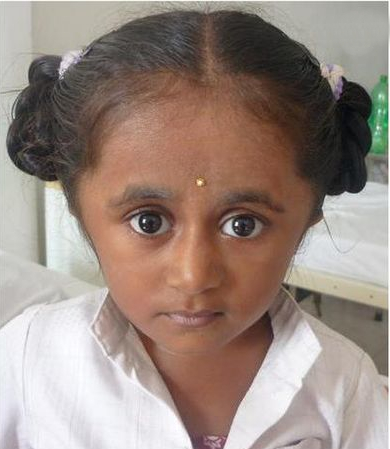
Other diagnostic factors
common
skin fragility
Patients with OI may have skin fragility and easy bruising. In one study of 959 adults with OI, 42% self-reported a history of bruising.[28]
easy bruising
Patients with OI may have skin fragility and easy bruising. In one study of 959 adults with OI, 42% self-reported a history of bruising.[28]
muscle weakness
Joint hypermobility and ligamentous laxity are common in OI. These manifest as chronic pain and increased muscle weakness/fatigability and can interfere with normal daily activities.
uncommon
cardiorespiratory signs and symptoms
Cardiorespiratory symptoms in patients with OI may include chest pain, palpitations, fatigue, syncope, dyspnoea, and cough. Patients may also present with signs of heart failure or arrhythmia.
More common in individuals with severe OI.
Risk factors
strong
family history of OI
For OI caused by pathogenic variants in COL1A1, COL1A2, and IFITM5, the mode of transmission is autosomal dominant.[8][9][10][25] Thus, an affected individual with OI will have a 50% chance of having an affected offspring. In recessive forms of OI, parents of affected individuals are obligate carriers. Thus, with each pregnancy, there is a 25% chance of having another affected child.
Use of this content is subject to our disclaimer