Images and videos
Images


Aortic dissection
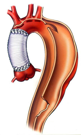
Dissection status post-proximal repair with late distal aneurysm
See this image in context in the following section/s:
Aortic dissection
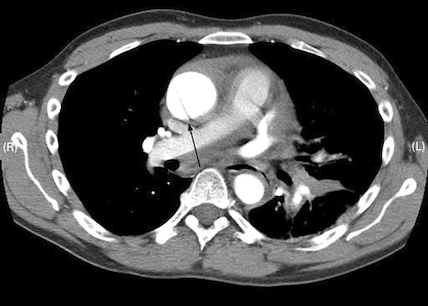
CT scan showing dissecting aneurysm in a 45-year-old patient with Marfan syndrome experiencing chest pain
See this image in context in the following section/s:

Aortic dissection
Multiparametric diagnostic work-up of acute aortic syndrome
Mazzolai L et. al, 2024 ESC guidelines for the management of peripheral arterial and aortic diseases: developed by the task force on the management of peripheral arterial and aortic diseases of the European Society of Cardiology (ESC) endorsed by the European Association for Cardio-Thoracic Surgery (EACTS), the European Reference Network on Rare Multisystemic Vascular Diseases (VASCERN), and the European Society of Vascular Medicine (ESVM), 45(36), 3538-700, https://doi.org/10.1093/eurheartj/ehae179. Reprinted by permission of Oxford University Press on behalf of the European Society of Cardiology. http://www.escardio.org/Guidelines
See this image in context in the following section/s:

Aortic dissection
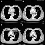
CT of a 71-year-old man showing type II dissecting aneurysm of the ascending aorta. Haematoma around the proximal segment of the ascending aorta (panels A-D) compressed the right pulmonary artery, almost occluding its patency and limiting the perfusion of the reciprocal lung
See this image in context in the following section/s:

Aortic dissection
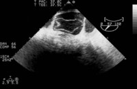
Trans-oesophageal echocardiography (transverse aortic section) showing a circumferential dissection of the ascending aorta in a 30-year-old patient with features of Marfan's syndrome
Bouzas-Mosquera A, Solla-Buceta M, Fojón-Polanco S. Circumferential aortic dissection. BMJ Case Reports 2009; doi:10.1136/bcr.2007.049908
See this image in context in the following section/s:
Use of this content is subject to our disclaimer