Approach
Following the exclusion of life-threatening conditions associated with urticaria (i.e., anaphylaxis and airway obstruction), the evaluation of patients with urticaria/angio-oedema often proceeds as follows:
Determine whether urticaria/angio-oedema is acute (<6 weeks) or chronic (≥6 weeks).
Perform a comprehensive history and physical examination to determine the most likely aetiology. A specific cause is more likely to be found in people with acute urticaria, while chronic urticaria is usually idiopathic in people with a normal history and physical examination.
Perform diagnostic testing, guided by abnormal/suspicious findings on history and physical examination.
History: acute versus chronic
In the case of acute urticaria/angio-oedema, an aetiology is more likely to be found and treated (or self-resolved in the case of viral infections) leading to resolution of disease within 6 weeks. If aetiology is missed then urticaria/angio-oedema may persist and become chronic. Patients with chronic urticaria/angio-oedema with a normal history and physical examination are likely to have idiopathic disease, also known as chronic spontaneous urticaria.[3] This accounts for approximately 80% to 90% of people with chronic urticaria/angio-oedema.[1][16]
History: possible aetiology
Because almost every disease state, from infections to endocrinopathies, has been associated with the pathogenesis of urticaria/angio-oedema, the initial history and physical examination should comprehensively look for the presence of any disease. Specific signs and symptoms will help point towards the aetiology. For instance, the aetiology for urticaria in patients with concomitant rhinorrhea and cough would likely be a viral infection.
History should take into consideration the following areas.
Medications and supplements
A major cause of urticaria/angio-oedema.
Usually elicited via an extensive medical history; however, given patients may have poor immediate recall, this may have to be elicited over several visits. Use of electronic medical records may help.
Ensure that questions cover health supplements, vitamins, and alternative medicine agents, as patients often think these are not 'medications'.
Food allergens
Usually, culprit foods are temporally related, with rashes occurring within minutes to hours of ingestion. Therefore, clinicians should focus on foods eaten close to time of urticaria/angio-oedema.
Certain foods may be hidden, such as soy used as fillers.
Food ingredients should be studied for a common agent present at the time of eruptions, especially in highly-processed meals.
A food and urticaria diary may help.
Infections
Viral infections are usually easily identified, especially in children.
Because occult infections have been implicated in urticaria/angio-oedema, a comprehensive screening history should be performed. Ask about dental, skin, gastrointestinal, and genitourinary infections, including bacterial and fungal aetiologies.
Asymptomatic infections, such as hepatitis, should be considered in people from high-risk populations (e.g., people of Asian origin).
Endocrinopathies
Any symptoms/signs of possible endocrine abnormalities, especially thyroid disease should be considered.
Malignancies
Any symptoms/signs of possible malignancy should be considered.
If there are no overt symptoms or signs of malignant transformation, age-appropriate malignancy screens should be performed (if not previously performed).
Autoimmune diseases
Any typical symptoms and signs of collagen vascular disease should be considered.
Autoimmune diseases often evolve over time.
When there are incomplete or non-specific symptoms and signs of specific syndromes (e.g., fatigue or arthralgias), diagnostic testing may be warranted, such as erythrocyte sedimentation rate, antinuclear antibodies, rheumatoid factor.
Physical urticarias
Pressure, vibration, solar/heat/cold/water exposure, and exercise can elicit urticarias. The history should be directed towards whether these physical factors seem to be associated with the development of urticaria/angio-oedema.
These aetiologies are common to the development of urticaria/angio-oedema but are not exhaustive. A full and comprehensive medical history is required, including thorough description of the presenting complaint, past medical history, family history, social history, and systems enquiry, and questions relating to quality of life and the emotional impact of the condition.[3]
Certain conditions may present with urticaria/angio-oedema but are not classified as subtypes of urticaria because they have quite different pathophysiological mechanisms.[3] Likewise, several syndromes that may feature wheals are not considered a subtype of urticaria, but related to it. These conditions need to be considered in the differential diagnosis and include:[28][29][30]
Cutaneous mastocytosis (urticaria pigmentosa)
Urticarial vasculitis
Auto-inflammatory syndromes (e.g., cryopyrin-associated periodic syndromes or Schnitzler’s syndrome)
Bradykinin-mediated angio-oedema (e.g., HAE)
Transfusion reactions
Serum sickness.
Other skin conditions with rashes that need to be differentiated from urticaria include:
Drug eruptions (note that drug allergy can result in urticaria but drug eruptions may present in morbilliform and more persistent rashes)
Insect bites (papular rash)
Viral exanthems
Atopic dermatitis
Contact dermatitis
Bullous pemphigoid
Erythema multiforme[Figure caption and citation for the preceding image starts]: Palmar target lesionsFrom the personal collection of Nanette Silverberg, MD [Citation ends].
Stevens Johnson syndrome[Figure caption and citation for the preceding image starts]: Stevens-Johnson syndrome: targetoid lesion and epidermal lossFrom the personal collection of Dr A. Kowal-Vern [Citation ends].

Auriculotemporal syndrome.
Physical examination
As such a wide range of disease states are associated with urticaria/angio-oedema, the physical examination should be complete and encompassing so as not to miss any signs of a potential aetiology. An extensive discussion of such examinations is beyond the scope of this review, and current guidelines should be consulted for more detail.[1][3] Important considerations when performing a physical examination in a patient with urticaria are as follows:
Skin examination
The characteristic skin lesions present as sudden onset pruritic whealing of the superficial dermis, generally lasting well under 24 hours.[6] Typically, urticarial lesions blanch fully on pressure or diascopy. No overlying flaking or scaling should be noted, as this is a dermal process. It is useful to ask the patient if they have any photos of the lesions if they are not present at the time of evaluation. Urticaria may be accompanied by angio-oedema, a swelling of the deeper dermis and tissues. This can result in a colorless, non-pitting induration of the extremities, lips, and genitals.[31]
Differentiating urticarial vasculitis/cutaneous mastocytosis
In a patient with typical urticaria, individual crops of urticaria should be pruritic, blanch, and resolve within 24 hours. They may reappear in a different location soon, but underlying skin should remain normal after resolution. Urticarial vasculitis (a form of cutaneous vasculitis characterised by inflammation of the small blood vessels) and cutaneous mastocytosis (a disease involving abnormally high numbers of mast cells in the skin) should be considered if the lesions have the following atypical features:
Persist for longer than 24 hours
Have more of a burning sensation or pain
Do not blanch with pressure on the skin
Leave a pigmented area after resolution.
In these instances, biopsy may be necessary and further diagnostic workup performed to detect possible associated systemic disease, such as rheumatological conditions and mastocytosis characterised by mast cell infiltration of one or more extra-cutaneous organs (systemic mastocytosis). Seek expert advice regarding appropriate investigations for the diagnosis of systemic mastocytosis.[12]
It is important to note that differentiating urticarial swellings from cutaneous mastocytosis may be difficult. Early onset mastocytosis with localised areas of mast cell aggregates in the skin may present with single urticarial lesions (mastocytomas), multiple lesions with overlying hyperpigmentation (urticaria pigmentosa), or diffuse mastocytosis.[12][13] Darier sign (urtication after pressure) is common in mastocytosis lesions.[11] Blistering can accompany mastocytosis of childhood. In adults, lesions are usually red to brown small papules or macules but can be accompanied by telangiectasias.
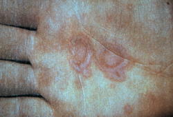
Dermatographism
This is whealing of skin minutes after a superficial sharp scratch. Dermatographism is found in 2% to 5% of the normal general population with no skin disease.[32][33][34][Figure caption and citation for the preceding image starts]: DermatographismWikipedia Commons; by JAguayo18 (CC BY-SA 4.0) [Citation ends].
 It is, however, associated with:
 Physical urticaria
Atopic dermatitis
Chronic spontaneous urticaria.
Investigations: acute urticaria
In most instances of acute urticaria/angio-oedema, a careful history and physical examination elicits diagnosis such that routine diagnostic testing is not recommended.[3] For instance, if peanuts were ingested 10 minutes before onset of rash and swelling, or a child develops signs and symptoms of an upper respiratory infection followed shortly by an urticarial rash, the history points towards the likely aetiology. In most of these patients, urticaria/angio-oedema resolves after resolution of the illness or avoidance of the allergen.
Diagnostic evaluation should be ordered in a targeted fashion, guided by abnormalities found on history and physical examination. For example, patients who have a suspected history of food-induced urticaria/angio-oedema may benefit from specific in vitro IgE or skin testing with the suspected culprit foods.
In general, in vitro and skin testing for food allergies have excellent negative predictive value, and negative tests generally exclude food allergy. Positive tests, unless extremely high in value, generally have a positive predictive value of approximately 50%.[35] They merely suggest the possibility of food allergy, which has to be confirmed with elimination of the food followed by challenge, with resolution and reappearance of urticaria respectively.
There are no validated tests for drug allergies, except for penicillin, and elimination of the suspected drug is often necessary for diagnosis of drug allergy.
Rapid streptococcal antigen testing may help in the evaluation of urticaria associated with symptoms of pharyngitis.
Referral to a specialist
Diagnostic testing is important in patients who have signs and symptoms suggestive of serious systemic illness, such as endocrinopathies, malignancy, and autoimmune disease, and referral to relevant specialists should be made. For instance, patients with urticaria as well as arthritis, oral sores, and alopecia should be tested for systemic lupus erythematosus (antinuclear antibodies [ANA], erythrocyte sedimentation rate [ESR], dsDNA etc), and referred to a rheumatologist. Routine testing for these pathologies among patients with urticaria along with a normal history and physical examination usually has a very low yield.
Referral to a dermatologist for skin biopsy is recommended for patients in whom lesions have abnormal characteristics (e.g., those that persist for longer than 24 hours, have a burning sensation, and leave pigmented areas) to include or exclude urticarial vasculitis or mastocytosis.
Investigations: chronic urticaria
Similar to acute urticaria, any diagnostic studies performed should be targeted to help confirm aetiology dependent upon findings on history and physical examination. Biopsy should be considered if urticaria lesions are atypical.
Approximately 80% to 90% of patients with chronic urticaria accompanied by a normal comprehensive history and physical examination have chronic spontaneous urticaria (formerly known as chronic idiopathic urticaria).[1][16] Of these, up to half may have an anti-IgE receptor antibody resulting in chronic release of mast cell mediators.[4][17][18][19] Therefore, a thorough history and physical examination may be sufficient to rule out exogenous aetiologies.
Only very limited routine diagnostic measures are recommended in chronic spontaneous urticaria.[3] These measures may not be necessary in some patients, but can be used to exclude underlying causes.[1]
These initial routine tests may include:
Complete blood count with differential
Serum chemistry, including liver function testing
ESR
Urinalysis
Thyroid studies.
The choice of investigations may vary by institution. Although these tests do not completely rule out all serious disease, they are a screen for chronic infection, parasites, endocrinopathies, autoimmune disease, and some malignancies, especially when they are used in conjunction with a thorough history and physical examination. Normal results will also provide reassurance to the patient who may require significant amounts of medication to suppress skin disease.
Assessment tools for chronic spontaneous urticaria include the Urticaria Activity Score (UAS7), which assesses the degree of pruritus and hive count over 7 days. It is a simple questionnaire with possible scores from 0 to 6 per day and from 0 to 42 per week. The resulting weekly score can be used to follow the disease activity and the efficacy of the ongoing therapy for chronic spontaneous urticaria.[36][37]
Physical (inducible) urticaria
Diagnostic tests are usually not needed for urticaria patients in which physical stimuli reliably elicit skin disease. Sometimes, when diagnosis is not apparent or response to treatment is inadequate, physical challenges may be performed.[4] These are best performed by clinicians or centers with expertise.
Pressure urticaria: eliciting urticaria in response to pressure after weight placement.
Solar urticaria: eliciting urticaria after exposure to light of various wavelengths and natural sunlight.
Vibratory urticaria: eliciting urticaria after exposure to a laboratory vortex mixer.
Aquagenic urticaria: eliciting urticaria after application of wet compress.
Cold-induced urticaria: eliciting urticaria after placement of ice cube.
Cholinergic urticaria: eliciting urticaria after raising core temperature and/or after intradermal injection of methacholine (high false negative rate).
Dermatographism: eliciting urticaria after scratching skin.
Special consideration of patients with angio-oedema without urticaria
Patients with angio-oedema without urticaria need to be screened for hereditary angio-oedema (HAE).[1] Measuring complement 4 (C4) levels is cost effective.
Patients with hereditary and acquired forms of angio-oedema, including consumption of C1 inhibitor from malignancy and autoimmune disease, universally have low C4 levels. Further measurement of C1 inhibitor quantity and function can be obtained if C4 is low. HAE can be classified broadly into two types: HAE due to C1INH deficiency (HAE C1INH) or HAE with normal C1INH (HAE-nl-C1INH).[26] Acquired angio-oedema from a paraneoplastic process results in abnormal C1q levels.
There are other forms of angio-oedema with absent urticaria with normal C1 inhibitor quality/function and normal C4 caused by over-expression of bradykinin. There are no specific tests for these patients, and diagnosis is usually confirmed with a trial of bradykinin inhibitor,. In general, these patients, especially those in whom swelling is severe or involving the airway, should be managed by clinicians with experience in this area.
European guideline approach to assessment of urticaria and angio-oedema
Guidance from the European Academy of Allergy and Clinical Immunology (EAACI), the EU-funded network of excellence, the Global Allergy and Asthma European Network (GA2 LEN), the European Dermatology Forum (EDF), and the World Allergy Organization (WAO) has been used to create a diagnostic algorithm for patients presenting with wheals, angio-oedema, or both.[3]
Healthcare professionals should elicit a thorough history, perform a physical examination, and conduct provocation tests (including for drugs, food) and physical tests as indicated by the history.[3]
Use of this content is subject to our disclaimer