Investigations
1st investigations to order
arthrocentesis with synovial fluid analysis
Test
Provides definitive diagnosis.[52][53] Excludes septic arthritis and differentiates gout from pseudogout (calcium pyrophosphate deposition disease).
The synovial fluid WBC count usually exceeds 2000/mm³, and the cells are mostly polymorphonuclear neutrophils type. Monosodium urate crystals (intracellular and/or extracellular needle-shaped crystals strongly negative for birefringence under polarised light) confirm the diagnosis.
Synovial fluid analysis should be considered in most patients, but the diagnosis can often be made clinically.
In the UK, the National Institute for Health and Care Excellence (NICE) recommends that arthrocentesis (with microscopy of synovial fluid) should be considered when the diagnosis of gout remains uncertain or unconfirmed following measurement of serum urate level.[52]
At times, poor transportation conditions or a long lag time between obtaining the synovial fluid and examining the specimen make it difficult to identify the crystals.
An expert, such as a rheumatologist or experienced technician, should examine the synovial fluid.
If the analysis fails to show monosodium urate crystals or other aetiology for the acute inflammatory arthritis, repeating arthrocentesis during future attacks should be considered.
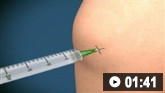
How to aspirate synovial fluid from the knee and administer intra-articular medication using a medial approach.
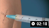
How to aspirate synovial fluid from the shoulder and administer intra-articular medication. Video demonstrates a posterior approach to the glenohumeral joint and a lateral approach to the subacromial space.
Result
WBC count >2.0 x 10⁹/L (2000/mm³ or 2000/microlitre; mean, 20,000/mm³ or 20,000/microlitre); strongly negative birefringent needle-shaped crystals under polarised light
Investigations to consider
serum uric acid level
Test
May be low, normal, or high during an acute gout attack. This test becomes more reliable when done at least 2 weeks after the attack resolves.[58] Gout can develop with levels lower than the upper limit of normal values.[58]
In the UK, NICE recommends performing a serum urate level as the first investigation to confirm the clinical diagnosis in patients with signs and symptoms of gout. A serum urate level 360 micromol/L (6 mg/dL) or more confirms a diagnosis of gout. If the serum urate level is below 360 micromol/L (6 mg/dL) during a gout flare, and gout is suspected, the test should be repeated at least two weeks after the flare has settled.[52]
Result
>420 micromol/L (>7 mg/dL) in men; >360 micromol/L (>6 mg/dL) in women
ultrasound
Test
Ultrasound-detected erosions are most commonly found in the first metatarsophalangeal joint and the metacarpophalangeal joints.[76]
Ultrasound findings, including tophi and erosion beside a double contour sign, have a sensitivity of 65% and specificity approaching 90%.[59][60]
Ultrasound is recommended for patients in the UK if joint aspiration can't be performed, or if the diagnosis of gout is uncertain.[52]
Result
erosions, tophi, double contour line
dual energy computed tomography (DECT)
Test
Could be helpful in the diagnosis of gout when it is in question, or for patients with contraindications for, or who refuse to have joint aspiration.[52][61][62][63]
Evidence suggests that DECT is valid and reliable, more sensitive than radiographs and CT, and at least comparable to ultrasound for the diagnosis of gout.[64][65][66]
Result
erosions, tophi, double contour line
x-ray of affected joint
Test
Radiographs are of limited diagnostic utility.[51] In late/severe gout, radiographic changes may help to differentiate between chronic gout and other joint conditions.[68]
X-ray findings suggestive of gout include soft-tissue opacifications with densities between soft tissue and bone, articular and periarticular bone erosions, and osteophytes at the margins of opacifications or erosions.[69]
The hands are an optimal place to look for gouty erosions.
Result
periarticular erosions (may have an overhanging edge or punched-out appearance)
Use of this content is subject to our disclaimer