History and exam
Key diagnostic factors
common
presence of risk factors
erythema and/or ulceration of oral mucosa
Oral mucosal changes range from erythema to patchy or confluent ulceration with a superficial pseudomembranous membrane or, rarely, overt necrosis.[2]
Lesions may be unilateral or bilateral, and are typically limited to non-keratinised oral mucosa. Common sites include the buccal mucosa, lip mucosa, ventral tongue, and soft palate.[Figure caption and citation for the preceding image starts]: Mucositis: 'buccal' mucosaFrom the teaching collection of Rajesh V. Lalla, DDS, PhD, CCRP, DABOM; used with permission [Citation ends].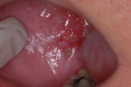
[Figure caption and citation for the preceding image starts]: Mucositis: dorsolateral tongueFrom the teaching collection of Rajesh V. Lalla, DDS, PhD, CCRP, DABOM; used with permission [Citation ends].
May be complicated by secondary viral or fungal infection.[1][2]
Other diagnostic factors
common
intra-oral bleeding
Bleeding may occur from ulcerative lesions in patients who are thrombocytopenic secondary to high-dose chemotherapy.[44]
dietary impairment and/or weight loss
Ability to maintain normal food intake and oral hydration is influenced by the severity of mucositis. It is important to assess the presence and extent of impairment (e.g., if the patient can eat a normal diet; eat and swallow a modified diet; or is unable to eat or drink adequately).
diarrhoea
May be a presenting feature in patients with associated gastrointestinal mucositis.[34]
nausea and/or vomiting
May be a presenting feature in patients with associated gastrointestinal mucositis.[34]
abdominal pain
May be a presenting feature in patients with associated gastrointestinal mucositis.[34]
uncommon
fever
Fever may be related to the presence of secondary infection, or be due to febrile neutropenia following chemotherapy.
See Febrile neutropenia.
Risk factors
strong
intensive chemotherapy regimens
Some chemotherapy drugs are associated with mucositis more than others.[4] Fluorouracil is a particularly mucotoxic drug. Regimens involving bolus dosing of fluorouracil are also more likely to cause mucositis than those involving infusion over longer periods of time. Additionally, chemotherapy regimens involving docetaxel plus doxorubicin plus cyclophosphamide (TAC) for breast cancer are known to confer a greater risk of oral mucositis.
Patients undergoing haematopoietic stem cell transplantation, particularly those receiving methotrexate, melphalan plus methotrexate, and BEAM (carmustine, etoposide, cytarabine, melphalan), are also at significant risk.[5][20]
radiotherapy to the oral cavity
Radiation toxicity is typically limited to the area included in the field of radiation. Therefore, oral mucositis is more common among patients with a primary malignancy involving the oral cavity or oropharynx, compared with those with a primary malignancy in other head and neck sites.[12] Severity of radiation mucositis is dose- and schedule-dependent. Most patients who receive greater than 50 Gy radiation develop ulcerative mucositis, and this condition is more likely in patients receiving altered fractionation schedules than in those receiving conventional radiotherapy.[6][13]
chemoradiation
Patients who receive chemotherapy concurrent with head and neck radiotherapy are significantly more likely to develop oral mucositis (OM) than those who do not.[13]
Additionally, patients undergoing haematopoietic stem cell transplantation and receiving total body irradiation, as part of myeloablative conditioning regimens, are at higher risk of developing severe OM.[20]
weak
genetic polymorphisms in drug metabolic enzymes
Polymorphisms in genes coding for enzymes involved in metabolism of chemotherapy agents can affect mucositis risk. For example, a mutation in dihydropyrimidine dehydrogenase, which metabolises fluorouracil, results in increased mucositis where fluorouracil is used in treatment.[21]
use of targeted therapies and immunotherapies
Use of this content is subject to our disclaimer