Images and videos
Images


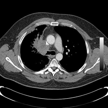
Central airway obstruction
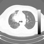
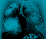
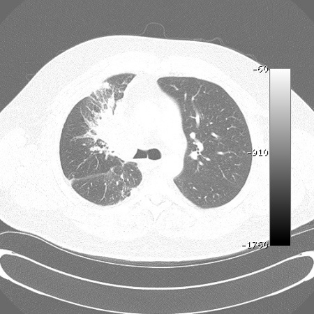
Malignant endobronchial obstruction on multidetector chest CT: lung window demonstrating right mainstem malignant obstruction
From the collections of Jose Fernando Santacruz MD, FCCP, DAABIP and Erik Folch MD, MSc; used with permission
See this image in context in the following section/s:
Central airway obstruction
Malignant endobronchial obstruction on multidetector chest CT: mediastinal window demonstrating right mainstem malignant obstruction
From the collections of Jose Fernando Santacruz MD, FCCP, DAABIP and Erik Folch MD, MSc; used with permission
See this image in context in the following section/s:

Central airway obstruction
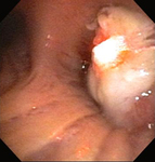
Bronchoscopic therapy for central airway obstruction of the right mainstem: laser photoresection
From the collections of Jose Fernando Santacruz MD, FCCP, DAABIP and Erik Folch MD, MSc; used with permission
See this image in context in the following section/s:
Central airway obstruction
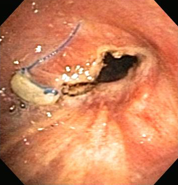
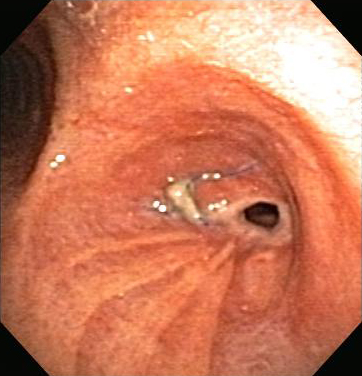
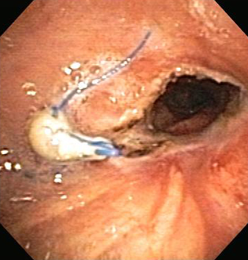
Post-lung transplant anastomotic bronchial stenosis: electrocautery radial incision
From the collections of Jose Fernando Santacruz MD, FCCP, DAABIP and Erik Folch MD, MSc; used with permission
See this image in context in the following section/s:
Central airway obstruction
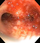
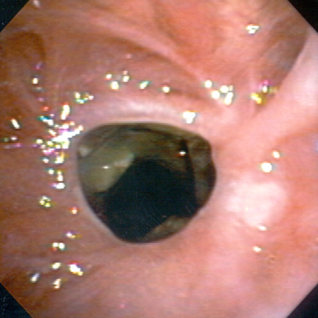
Airway stenosis secondary to granulomatosis with polyangiitis (formerly known as Wegener's granulomatosis)
From the collections of Jose Fernando Santacruz MD, FCCP, DAABIP and Erik Folch MD, MSc; used with permission
See this image in context in the following section/s:
Central airway obstruction
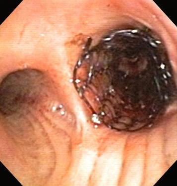
Central airway obstruction: malignant obstruction of the right mainstem
From the collections of Jose Fernando Santacruz MD, FCCP, DAABIP and Erik Folch MD, MSc; used with permission
See this image in context in the following section/s:
Central airway obstruction
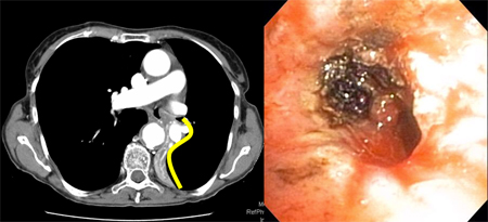
Golden-S or reverse S-sign on chest CT (left image) traced by yellow line. Flexible bronchoscopy (right image) shows the central obstructive lesion at the left mainstem bronchi.
From the collections of Jose Fernando Santacruz MD, FCCP, DAABIP and Erik Folch MD, MSc; used with permission
See this image in context in the following section/s:
Central airway obstruction
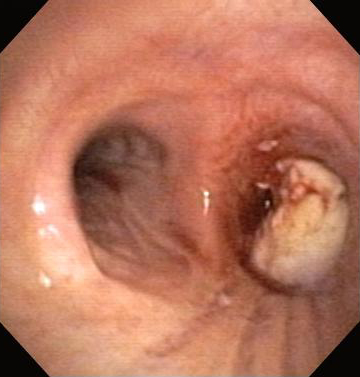
Post-lung transplant anastomotic bronchial stenosis
From the collections of Jose Fernando Santacruz MD, FCCP, DAABIP and Erik Folch MD, MSc; used with permission
See this image in context in the following section/s:
Central airway obstruction
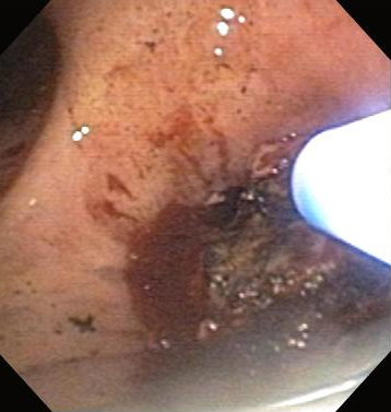
Bronchoscopic therapy for central airway obstruction of the right mainstem: argon plasma coagulation
From the collections of Jose Fernando Santacruz MD, FCCP, DAABIP and Erik Folch MD, MSc; used with permission
See this image in context in the following section/s:
Central airway obstruction
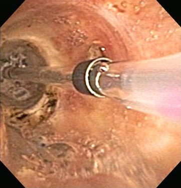
Post-lung transplant anastomotic bronchial stenosis: balloon bronchoplasty
From the collections of Jose Fernando Santacruz MD, FCCP, DAABIP and Erik Folch MD, MSc; used with permission
See this image in context in the following section/s:
Central airway obstruction
Post-lung transplant anastomotic bronchial stenosis: right mainstem anastomosis post-multimodal endoscopic therapy
From the collections of Jose Fernando Santacruz MD, FCCP, DAABIP and Erik Folch MD, MSc; used with permission
See this image in context in the following section/s:
Central airway obstruction
Central airway obstruction: malignant obstruction of the right mainstem
From the collections of Jose Fernando Santacruz MD, FCCP, DAABIP and Erik Folch MD, MSc; used with permission
See this image in context in the following section/s:

Central airway obstruction
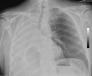
Chest x-ray showing right mainstem endobronchial stent occlusion with mucus
From the collections of Jose Fernando Santacruz MD, FCCP, DAABIP and Erik Folch MD, MSc; used with permission
See this image in context in the following section/s:
Central airway obstruction
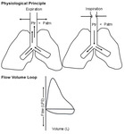
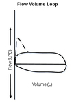
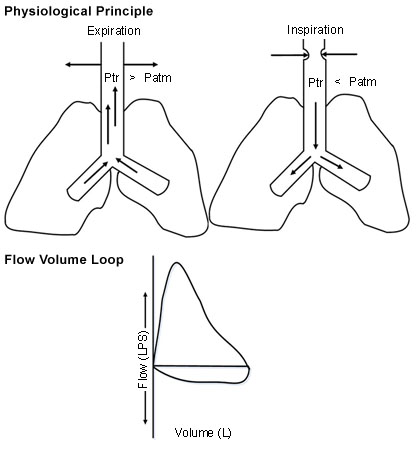
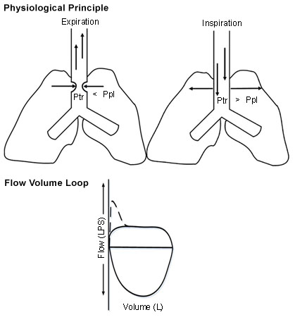
Dynamic (non-fixed or variable) extrathoracic upper airway obstruction: flow-volume loop shows 'flattened' inspiratory limb during forced inspiration
From the collections of Jose Fernando Santacruz MD, FCCP, DAABIP and Erik Folch MD, MSc; used with permission
See this image in context in the following section/s:
Central airway obstruction
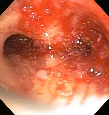
Bronchoscopic therapy for central airway obstruction of the right mainstem: post-mechanical debulking
From the collections of Jose Fernando Santacruz MD, FCCP, DAABIP and Erik Folch MD, MSc; used with permission
See this image in context in the following section/s:
Central airway obstruction
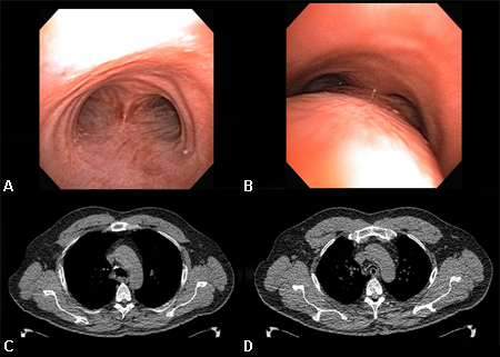
Dynamic airway collapse: A. bronchoscopic view on inhalation; B. bronchoscopic view on exhalation showing dynamic airway collapse; C. CT chest showing normal airway on inhalation; D. CT chest showing significant airway collapse on exhalation
From the collections of Jose Fernando Santacruz MD, FCCP, DAABIP and Erik Folch MD, MSc; used with permission
See this image in context in the following section/s:
Central airway obstruction
Bronchoscopic therapy for central airway obstruction of the right mainstem: stent placement
From the collections of Jose Fernando Santacruz MD, FCCP, DAABIP and Erik Folch MD, MSc; used with permission
See this image in context in the following section/s:
Central airway obstruction
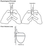
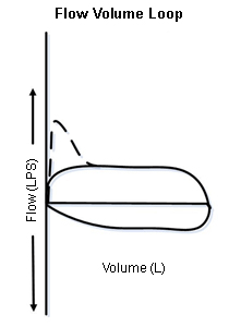
Dynamic (non-fixed or variable) intrathoracic upper airway obstruction: flow-volume loop shows 'flattened' expiratory limb during forced exhalation
From the collections of Jose Fernando Santacruz MD, FCCP, DAABIP and Erik Folch MD, MSc; used with permission
See this image in context in the following section/s:

Central airway obstruction
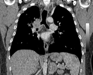
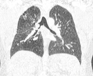
Malignant endobronchial obstruction on multidetector chest CT: coronal reconstruction demonstrating right mainstem malignant obstruction
From the collections of Jose Fernando Santacruz MD, FCCP, DAABIP and Erik Folch MD, MSc; used with permission
See this image in context in the following section/s:
Central airway obstruction
Chest x-ray showing right mainstem endobronchial stent occlusion with mucus
From the collections of Jose Fernando Santacruz MD, FCCP, DAABIP and Erik Folch MD, MSc; used with permission
See this image in context in the following section/s:

Central airway obstruction
Malignant endobronchial obstruction on multidetector chest CT: 2D multiplanar reconstruction (minimal intensity projection) demonstrating right mainstem malignant obstruction
From the collections of Jose Fernando Santacruz MD, FCCP, DAABIP and Erik Folch MD, MSc; used with permission
See this image in context in the following section/s:
Central airway obstruction
Fixed upper airway obstruction: flow-volume loop shows 'flattened' inspiratory and expiratory loops
From the collections of Jose Fernando Santacruz MD, FCCP, DAABIP and Erik Folch MD, MSc; used with permission
See this image in context in the following section/s:

Central airway obstruction
Malignant endobronchial obstruction on multidetector chest CT: 3D volume rendering reconstruction demonstrating right mainstem malignant obstruction
From the collections of Jose Fernando Santacruz MD, FCCP, DAABIP and Erik Folch MD, MSc; used with permission
See this image in context in the following section/s:
Central airway obstruction
Post-lung transplant anastomotic bronchial stenosis
From the collections of Jose Fernando Santacruz MD, FCCP, DAABIP and Erik Folch MD, MSc; used with permission
See this image in context in the following section/s:
Central airway obstruction
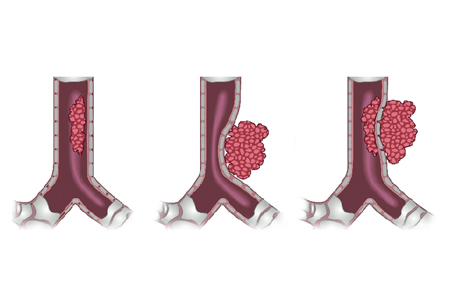
Types of central airway obstruction: intrinsic, extrinsic, and mixed
From the collections of Jose Fernando Santacruz MD, FCCP, DAABIP and Erik Folch MD, MSc; used with permission
See this image in context in the following section/s:

Central airway obstruction
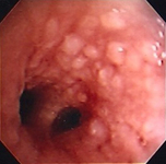
Tracheal-bronchial papillomatosis
From the collections of Jose Fernando Santacruz MD, FCCP, DAABIP and Erik Folch MD, MSc; used with permission
See this image in context in the following section/s:
Use of this content is subject to our disclaimer