Case history
Case history #1
A 55-year-old woman presents with a right flank mass. She states she was recently diagnosed with diabetes mellitus, which she has been able to control with diet modifications. She lost 9 kg (20 pounds) within 3 months and then noticed a mass over her right lower rib cage. She denies pain but does report discomfort when she wears a jogging bra. On physical examination, the mass is soft, superficial, and mobile, and it measures 5 cm in diameter.
Case history #2
A 35-year-old man presents with a right thigh nodule and a recurrent left chest wall nodule at the site of a prior scar. He states that he noticed a bump on his right lateral thigh 2 years previously and that the left chest wall lesion had been removed in clinic 3 years prior. The nodules have grown slightly over recent months. He also states that they bother him when he touches them. On physical examination, the nodules are 1 cm x 2 cm, soft, and mobile, and they feel subcutaneous.
Other presentations
Lipomas can present in locations other than subcutaneously on the trunk or proximal extremities. Gastrointestinal lipomas are uncommon and occur as submucosal lesions, most commonly in the stomach, small intestine, and colon.[7] This type may present with intestinal obstruction or bleeding. Rarely, lipomas can also occur in locations such as the adrenal glands, parotid glands, parapharyngeal space, breast, mediastinum, pleura, major airway, heart, superior vena cava, brain, and intraspinal areas.[8]
Lipomas can occur on a hereditary basis in patients with familial multiple lipomatosis.[9][10] Patients with this autosomal condition tend to be male and have multiple, widespread, symmetric lipomas of the extremities and trunk.[11] Other hereditary syndromes that involve lipomas include Madelung's disease, also known as multiple symmetric lipomatosis; Dercum's disease, also known as adiposis dolorosa; and Gardner's syndrome.[12] Madelung's disease is more common in men and is associated with chronic alcohol consumption in genetically predisposed individuals. Features include benign symmetric lipomatosis of the head, neck, shoulders, and proximal upper extremities.[13] Dercum's disease occurs in middle-aged women and is characterised by painful lipomas on the trunk, shoulders, arms, and legs.[14]
Angiolipomas account for approximately 10% of all lipomatous lesions.[15] They present as painful, subcutaneous nodules, usually in young adults, and are multiple in more than 50% of cases.[2][16] Angiolipomas are composed of adipocytes interspersed with clusters of capillaries containing fibrin thrombi.
Spindle cell lipomas, often seen in men between the ages of 45 and 65 years, occur in the posterior neck and shoulder area.[16] They are characterised by mature fat being replaced by collagen-forming spindle cells.[17][18]
Intramuscular lipomas, which are usually poorly circumscribed and infiltrative, typically present in mid-adult life as slow-growing, deep masses located in the thigh or trunk. It is important to exclude an atypical lipomatous tumour or well-differentiated liposarcoma, as these are more common than an intramuscular lipoma in this anatomical position.[16][19] Retroperitoneal lipomas are very rare and a diagnosis of atypical lipomatous tumour or well-differentiated liposarcoma should also be considered in patients with retroperitoneal lesions.[20]
Hibernomas may arise in the trunk, retroperitoneum, and extremities and resemble the glandular brown fat found in hibernating animals.[4][16] They have a greater tendency to bleed during excision and recur if not fully excised.[Figure caption and citation for the preceding image starts]: Subcutaneous lipoma on the trunkFrom the collection of Dr Kimberly Moore Dalal and Dr Steven D. DeMartini; used with permission [Citation ends].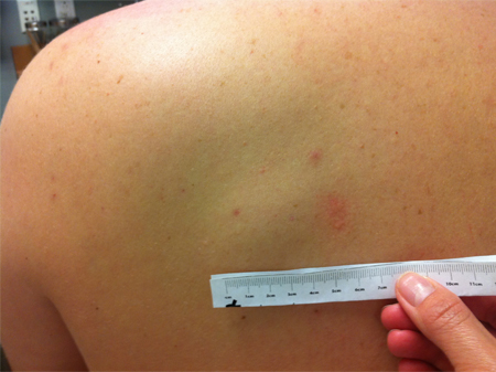 [Figure caption and citation for the preceding image starts]: Gastric submucosal lipoma, CT scan. Submucosal antral mass with fatty density throughout.From the collection of Dr Kimberly Moore Dalal and Dr Steven D. DeMartini; used with permission [Citation ends].
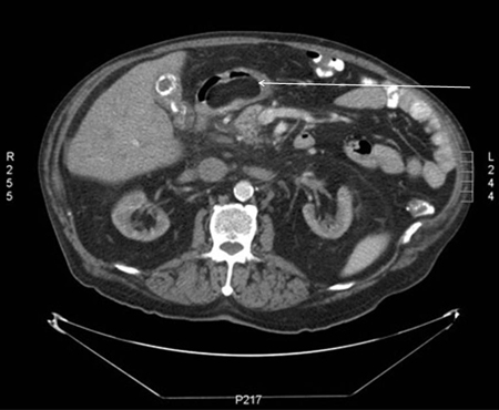 [Figure caption and citation for the preceding image starts]: Gastric submucosal lipoma, upper GI contrast study. Filling defect in the distal antrum and pyloric channel suggesting antral mass prolapsing into pyloric channelFrom the collection of Dr Kimberly Moore Dalal and Dr Steven D. DeMartini; used with permission [Citation ends].
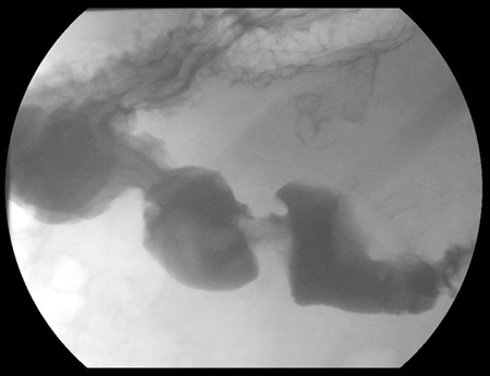 [Figure caption and citation for the preceding image starts]: Gastric submucosal lipoma. A nodule of mature adipose tissue is present subjacent to gastric mucosa. Haematoxylin and eosin, 20x magnificationFrom the collection of Dr Kimberly Moore Dalal and Dr Steven D. DeMartini; used with permission [Citation ends].
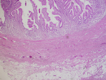 [Figure caption and citation for the preceding image starts]: Angiolipoma. Mature adipose tissue with foci of endothelial proliferation containing micro-vascular thrombi. Haematoxylin and eosin, 200x magnificationFrom the collection of Dr Kimberly Moore Dalal and Dr Steven D. DeMartini; used with permission [Citation ends].
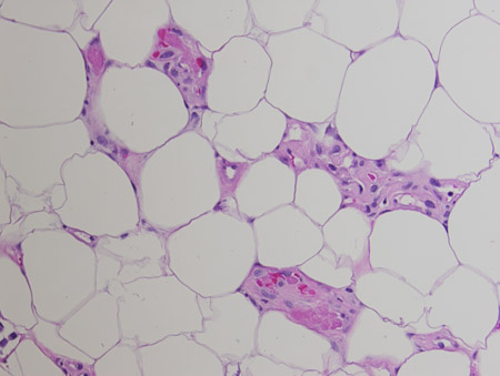 [Figure caption and citation for the preceding image starts]: Spindle cell lipoma. Mature adipose tissue with intervening strands of dense fibrosis with spindle cell areas and characteristic ropey collagen bundles. Haematoxylin and eosin, 200x magnificationFrom the collection of Dr Kimberly Moore Dalal and Dr Steven D. DeMartini; used with permission [Citation ends].
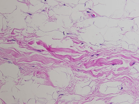 [Figure caption and citation for the preceding image starts]: Intramuscular lipoma, right thigh. MRI, axial, T1-weighted image. Lipomatous mass in the anterior aspect of the right thighFrom the collection of Dr Kimberly Moore Dalal and Dr Steven D. DeMartini; used with permission [Citation ends].
 [Figure caption and citation for the preceding image starts]: Intramuscular lipoma, right thigh. MRI, coronal, T1-weighted image. Lipomatous mass in the anterior aspect of the right thighFrom the collection of Dr Kimberly Moore Dalal and Dr Steven D. DeMartini; used with permission [Citation ends].
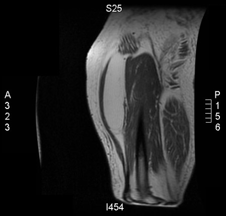 [Figure caption and citation for the preceding image starts]: Intramuscular lipoma of subscapularis muscle, CT scan. Right axillary soft-tissue fatty mass with well-circumscribed marginsFrom the collection of Dr Kimberly Moore Dalal and Dr Steven D. DeMartini; used with permission [Citation ends].
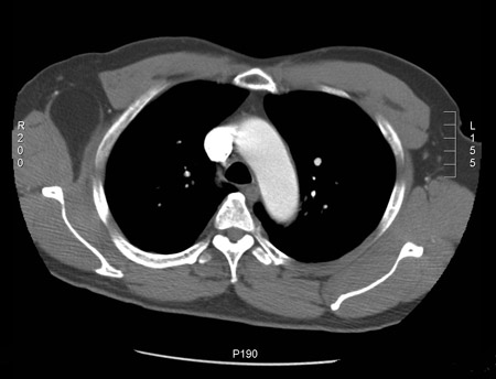 [Figure caption and citation for the preceding image starts]: Intramuscular lipoma. Mature adipose tissue insinuating between skeletal muscle bundles. Haematoxylin and eosin, 200x magnificationFrom the collection of Dr Kimberly Moore Dalal and Dr Steven D. DeMartini; used with permission [Citation ends].
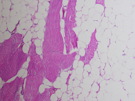
Use of this content is subject to our disclaimer