Investigations
Your Organisational Guidance
ebpracticenet urges you to prioritise the following organisational guidance:
Aspecifieke nekpijn: diagnose en behandelingPublished by: KCELast published: 2013Douleurs cervicales aspécifiques : diagnostic et traitementPublished by: KCELast published: 20131st investigations to order
cervical MRI
Test
Ordered if neck pain persists for 4 to 6 weeks, radicular pain does not subside with treatments, or more severe deficit suggestive of myelopathy is present. This would normally be the primary study ordered from the clinic setting once these criteria are met.[32][39]
MRI is also indicated for patients with neck pain and a history of malignancy, prior cervical spine surgery, or if there is suspicion for infection.[32]
[Figure caption and citation for the preceding image starts]: Severe, multi-level degenerative disc disease changes but without significant spinal cord compression (i.e., neither deformation nor intrinsic T2 changes) on cervical MRI (sagittal T2)Dennis A. Turner, MA, MD [Citation ends]. [Figure caption and citation for the preceding image starts]: Cervical MRI (sagittal T2) with mild degenerative joint disease and disc bulgingDennis A. Turner, MA, MD [Citation ends].
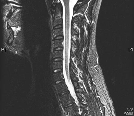 [Figure caption and citation for the preceding image starts]: Cervical MRI (sagittal T2) with moderate degenerative joint disease but no significant spinal cord compressionDennis A. Turner, MA, MD [Citation ends].
 [Figure caption and citation for the preceding image starts]: A single level of spinal cord compression with T2 changes, on cervical sagittal T2 sequence in the presence of symptomatic degenerative cervical myelopathyDennis A. Turner, MA, MD [Citation ends].
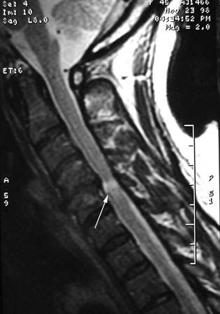 [Figure caption and citation for the preceding image starts]: Previous spinal cord compression at C3/4 on sagittal T2 MRI, with residual T2 changes, and new compression at C2/3 and C6/7, with T2 changesDennis A. Turner, MA, MD [Citation ends].
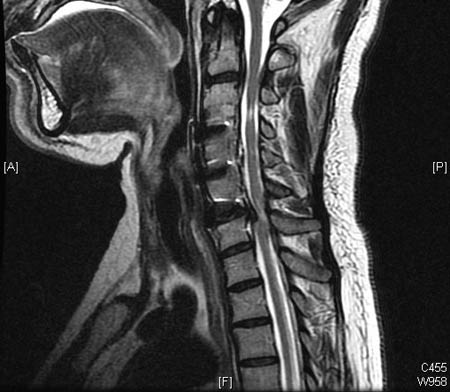
Result
bone destruction, spinal cord or nerve compression, intradural or epidural pathology
cervical x-ray
Test
Indicated for patients with prior cervical spin surgery with acute or increasing mechanical cervical pain or radiculopathy, and no trauma or red flags.[32]
May be appropriate for patients with acute or increasing cervical pain with or without radiculopathy and no trauma or red flags.[32]
Flexion/extension radiographs have limited value in degenerative disease.[40]
Result
presence of degenerative joint disease or degenerative disc disease, malalignment, spinal canal stenosis, fracture, or instability
Investigations to consider
cervical CT scan
Test
An extension to cervical radiographs to obtain more detail about bone structure, such as in ossified posterior longitudinal ligament calcification, trauma setting, or instability.
Also indicated if an MRI is not possible (e.g., implanted metal).[32]
Result
bone destruction, spinal cord or nerve compression; intradural or epidural pathology
cervical CT myelogram
Test
If a cervical CT scan with no contrast suggests spinal cord abnormalities and an MRI is not possible, then the next step is a CT cervical spine scan with intrathecal contrast (CT myelography) to obtain more detail about spinal cord and nerve changes.[32]
Primarily indicated only if an MRI is not possible (e.g., implanted metal).[32]
Result
presence of disc herniation or nerve root compression
cervical/upper extremity electromyography or nerve conduction velocity
Test
Ordered if brachial plexopathy, peripheral neuropathy, or peripheral nerve compression suspected, or mimicking radiculopathy or myelopathy.
Electromyography or nerve conduction velocity is a negative diagnostic test in cervical myelopathy, as nerve studies primarily demonstrate lower motor neuron changes. In degenerative cervical myelopathy, the neurological abnormalities arise from spinal cord compression and upper motor neuron dysfunction.
Result
may show changes of muscle denervation; localises the problem to the root or reveals changes in keeping with peripheral nerve entrapment
cervical nerve root block
Test
Ordered when specific nerve root involved with radiculopathy cannot be clinically or radiologically determined.[34]
Result
positive test if nerve block relieves patient's radicular pain temporarily; localises single nerve root
Use of this content is subject to our disclaimer