Case history
Case history #1
A 23-year-old woman presents with diarrhoea lasting several weeks. She has lost weight, but has not had a fever, and has not noticed blood in the stool. The diarrhoea started while she was travelling in Mexico.
Case history #2
A 39-year-old man presents to the accident and emergency department with right shoulder pain that he has been experiencing for 2 months. He reports that the pain has radiated to his back. He has been having night sweats and chills and has lost around 5 kg in weight. He has also had abdominal pain for 3 days and a non-productive cough. He was born in Iran and recently emigrated to the US. Physical examination identifies hepatomegaly and decreased breath sounds over the lower two-thirds of the right lung. Neurological examination is normal.
Other presentations
Amoebiasis is asymptomatic in around 80% to 90% of cases.[7] Diarrhoea is the most common illness caused by Entamoeba histolytica, although intestinal disease may present as dysentery (diarrhoea with blood or mucus) rather than diarrhoea alone. An amoeboma, which is a mass of granulation tissue in the colon that can be similar in appearance to colonic carcinoma, may also be detected.
Less common extraintestinal manifestations are peritonitis from perforation of the intestine, which sometimes mimics acute appendicitis, and pleural or pericardial effusions from direct extension of a liver abscess and brain abscess (almost all patients with brain abscess due to E histolytica also have a liver abscess).[1][2][3][4][5][6][8][9] Amoebic acute appendicitis is also a possible but rare manifestation of amoebiasis; amoebic appendicitis is more likely to be complicated than non-amoebic appendicitis.[9]
Rarely, clinical presentation may occur some years after primary infection.[7]
[Figure caption and citation for the preceding image starts]: Cyst of Entamoeba histolytica: unstained (A), and iodine stained (B) after formalin-ether concentration of stool sample.Original photos from National Center for Global Health and Medicine, Tokyo, Japan. [Citation ends].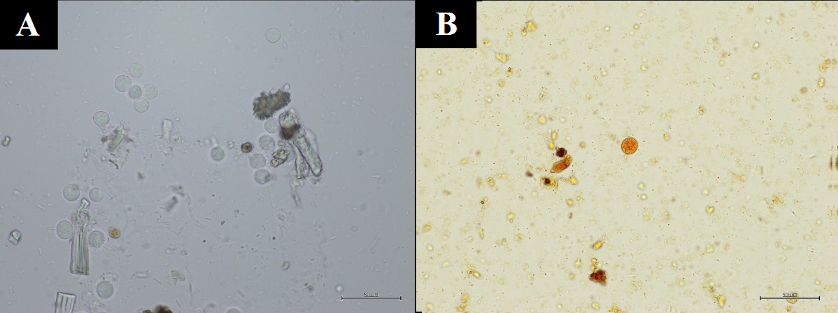 [Figure caption and citation for the preceding image starts]: Amoebic appendicitis with skin fistula two weeks after appendectomy (enhanced computed tomography).Original photo from National Center for Global Health and Medicine, Tokyo, Japan. [Citation ends].
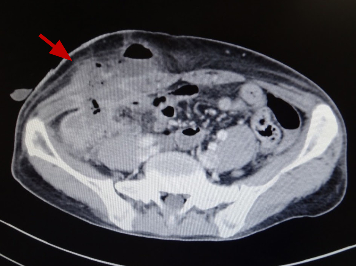 [Figure caption and citation for the preceding image starts]: Hematoxilin-Eosin stain (A-C) and Periodic acid-Schiff stain (D-F) of resected appendix of amoebic appendicitis. Entamoebas are deeply dyed by Periodic acid-Schiff stain.Original photo from National Center for Global Health and Medicine, Tokyo, Japan. [Citation ends].
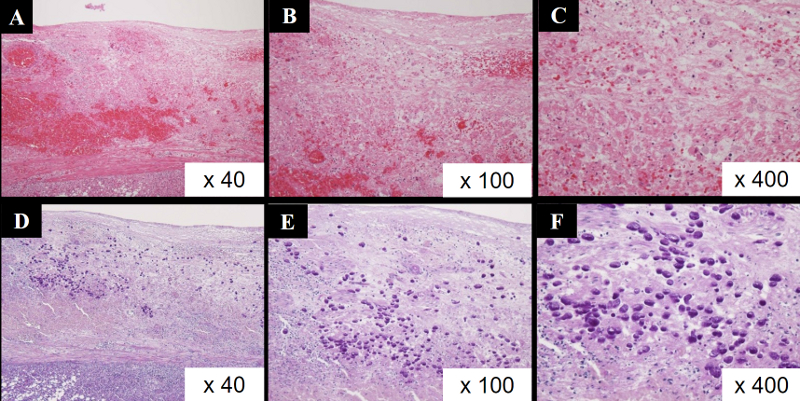
Use of this content is subject to our disclaimer