Approach
Respiratory alkalosis may produce a variety of clinical manifestations, ranging from mild symptoms to fatal outcome, due to direct effects or secondary electrolyte disturbances.
Respiratory alkalosis can be classified clinically into 3 categories:[3]
As a component of disease processes
Induced accidentally
Induced deliberately (therapeutic).
Accidental respiratory alkalosis develops as a consequence of inappropriate settings of mechanical ventilation or associated with extracorporeal membrane oxygenation (ECMO).[3]
History
Official consensus statements or guidelines on how to evaluate respiratory alkalosis are not available. However, brief inquiry into the following areas is almost always useful in establishing the cause.
Dyspnoea:
with pleuritic chest pain suggests pulmonary embolism (PE), pneumonia, or pneumothorax
with wheezing suggests asthma
with exertion may be the presenting signs of pulmonary oedema or pulmonary hypertension.
Productive cough and fever may suggest pneumonia. Cough may also be present in ARDS and interstitial fibrosis. Hemoptysis may be present with PE leading to infarction.
Fever itself should be noted because it may be a manifestation of systemic inflammatory-response syndrome (SIRS) or sepsis (although temperature should not be used as the sole predictor of sepsis and should not be used to rule sepsis either in or out).[119] When fever is associated with headache, stiff neck, and lethargy, it may signify meningitis. Meningeal symptoms with focal neurological complaints or signs suggest meningoencephalitis. Focal neurological symptoms may also signify CVA or a space-occupying lesion. Anxiety, pain, or a psychiatric history may point towards hyperventilation syndrome.
Risk factors for central sleep apnoea, such as CHF or CVA, should be elicited. Factors for liver disease, such as alcohol dependence and history of viral hepatitis or cirrhosis, should be noted. In a female patient of childbearing age, pregnancy needs to be considered. Medication history is important, particularly with regard to hormone-replacement therapy or salicylate overdose.
Obvious causes of acute tissue hypoxia include hypovolaemic, cardiogenic, or septic shock. Chronic causes of hypoxia, such as severe anaemia or haemoglobinopathy, should be sought. The clinician should be alert for conditions associated with hypoxaemia, such as ascent to high altitude, parenchymal lung disease, cyanotic heart disease, or portopulmonary hypertension.
Physical examination
Vital signs, including respiratory rate, temperature, blood pressure, heart rate, and pulse oximetry, should be obtained and monitored. Tachycardia may be present in sepsis, PE, or salicylate toxicity, or with pain. Lethargy or somnolence is present in meningitis or encephalitis. Scleral icterus and jaundice suggest hepatic failure.
Abnormalities in heart sounds or rhythm may reveal heart failure or valve abnormalities. A heart murmur and irregularly irregular rhythm may point towards embolic stroke. A loud pulmonic sound suggests pulmonary hypertension. Jugular venous distension may also be present in heart failure or pulmonary hypertension.
Unilateral hyper-resonance to percussion and decreased breath sounds suggest pneumothorax. Wheezing suggests bronchoconstriction. Crackles are present in pulmonary oedema, pneumonia, ARDS, and interstitial fibrosis.
Ascites, hepatomegaly, and caput medusae are present in liver failure or cirrhosis. A prominent uterus should lead one to suspect pregnancy in a woman of childbearing age.
Altered mental status may be present with CVA, meningoencephalitis, sepsis, or hepatic failure. Focal neurological signs may be present in CVA, encephalitis, and space-occupying brain lesions. Headache, neck stiffness, and meningeal signs are present in meningitis.
Arterial blood-gas analysis
Determination of respiratory alkalosis requires arterial blood gas analysis.

How to obtain an arterial blood sample from the radial artery.
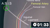
How to perform a femoral artery puncture to collect a sample of arterial blood.
When arterial blood cannot be obtained, there is a generally acceptable clinical correlation between arterial and venous blood pH and PCO₂ measurements. Based on Bland-Altman plots comparing venous and arterial blood samples, extrapolated arterial PCO₂ is calculated as follows:[128]
PCO₂ = (venous PCO₂ x 0.68) + 0.39
A small study involving critically ill patients compared the diagnostic accuracy of venous blood obtained from a central venous catheter with arterial blood samples. It revealed a diagnostic sensitivity of 100% in metabolic acidosis, metabolic alkalosis, and respiratory acidosis. However, sensitivity was only 71% in diagnosing respiratory alkalosis. Overall agreement was 69.6% between arterial and venous samples.[129]
Initial efforts are aimed at first determining whether respiratory alkalosis is acute:
change in HCO₃- (mmol/L) = 0.1 x change in PaCO₂ (mmHg)
or chronic:
change in HCO₃- (mmol/L) = 0.4 x change in PaCO₂ (mmHg)
When complemented with history, physical examination, and clinical data, these results guide detection of the underlying cause with special attention to urgent considerations. For instance, if respiratory alkalosis is acute, the focus is on signs and symptoms within the immediate prior 6 hours (e.g., PE or salicylate toxicity). If the disorder is chronic, attention is directed towards hormonal aetiologies (e.g., pregnancy, menstrual cycle, or hormone-replacement therapy), liver disease, or other chronic causes of hypoxia. It should be noted here that hyperventilation syndrome is a diagnosis of exclusion.
The arterial blood gas confirms and may offer all the clues necessary to discern the aetiology of respiratory alkalosis. A wide alveolar-arterial PO₂ gradient suggests disorders with ventilation-perfusion mismatch (e.g., pneumonia, PE) or shunt (e.g., cyanotic heart disease). A concurrent anion gap metabolic acidosis suggests salicylate poisoning. Concurrent wide alveolar-arterial PO₂ gradient with anion gap metabolic acidosis suggests tissue hypoxia, such as hypovolaemic, septic, or cardiogenic shock, severe anaemia, or haemoglobinopathy.
Chest radiograph
In addition to arterial blood-gas analysis, a chest radiograph should be obtained. A chest radiograph will identify pneumothorax and pulmonary parenchymal aetiologies of respiratory alkalosis. It should be noted; however, that the chest radiograph is a poor tool to detect PE, early ARDS, early fibrotic lung disease, and pulmonary hypertension.
Brain imaging
In patients with a suspected neurological aetiology, brain imaging should be obtained. Head CT is effective in evaluating for haemorrhage and mass effect; however, MRI may be needed for ischaemic stroke. Brain imaging may aid in the diagnosis of meningitis and encephalitis, but lumbar puncture should be performed if these diagnoses are being considered.
Other tests
If the chest radiograph is unremarkable, and PE remains a high probability, CT angiography of the pulmonary arteries is necessary. Ventilation/perfusion (V/Q scan) and echocardiography also are helpful adjuncts in this regard. Pulmonary function tests may be useful in chronic pulmonary diseases such as pulmonary fibrosis or asthma. Technetium-labelled macroaggregated albumin can assist in the diagnosis of intrapulmonary shunt. Echocardiogram and right heart catheterisation should be performed in patients with suspected pulmonary hypertension from any cause. Serum drug screen for salicylates or other stimulants is often useful as well. In summary, diagnostic tests should be based on the clinical suspicion of specific conditions.
Use of this content is subject to our disclaimer