Investigations
1st investigations to order
serology
Test
Enzyme-linked immunosorbent assay may be run on serum or cerebrospinal fluid for detection of Hendra virus (HeV)- or Nipah virus (NiV)-specific antibodies.
May be negative very early in the course of the illness. For NiV, IgM has been reported to be positive in two-thirds of cases by day 4 and 100% of cases by day 12, whereas it takes until day 26 for 100% of cases to be IgG positive.[36][42] IgG appears to remain positive persistently, whereas IgM starts to become undetectable after 3 months (although it can persist for longer than 7 months in some patients).[43][42]
By contrast, diagnostic sensitivity of serological tests is less well established in HeV.[36]
The degree of cross-reactivity with other infections is not yet firmly established.
It is recommended to seek advice from local virology services as to best samples/assays and transportation arrangements.
Result
positive IgM by 1-2 weeks; positive IgG by 2-3 weeks
reverse transcription-polymerase chain reaction
Test
Detection of viral RNA from serum, urine, respiratory, and/or cerebrospinal fluid samples.[36]
Most useful early in the course of illness.
Need to obtain advice from local virology services as to whether available locally or nationally. Yield and positive and negative predictive values presently unknown.
Result
positive for NiV or HeV RNA
FBC
Test
Thrombocytopenia can occur in both Hendra virus (HeV) and Nipah virus (NiV).[22][14][29] Leukopenia may be seen in NiV.[22] Neutropenia has been reported in HeV infection.[29]However, FBC may be normal, and many other infections produce a low platelet count and/or abnormal leukocyte counts.
Result
may be normal; commonly shows thrombocytopenia; leukopenia or neutropenia may also be seen
LFTs
serum urea and electrolytes
Test
A small number of patients developed syndrome of inappropriate ADH in their recovery phase in the Malaysian NiV outbreak.[14] Hyponatraemia is, however, not specific to NiV.
Result
may be normal or show hyponatraemia
clotting profiles
Test
Changes are not pathognomonic or specific for henipavirus infections. However, clotting profile is an important test for general clinical care.
Result
may be normal
CXR
Test
Changes are not pathognomonic or specific for henipavirus infections.
Consolidation and/or reticular changes may be seen, as well as diffuse bilateral opacities consistent with acute respiratory distress syndrome.[14][37][38] More CXR changes were noted in the NiV outbreaks occurring in Bangladesh compared with Malaysia, possibly owing to strain differences.
Less is known about CXR changes with HeV infection, but they may be normal on presentation.[29]
Result
may be normal or show focal consolidation, reticular changes, or diffuse infiltrates
malaria thick and thin blood films
Test
A negative result excludes malaria.
Result
negative
blood/urine cultures
Test
A negative result excludes other potential aetiologies.
Result
negative
cerebrospinal fluid analysis
cerebrospinal fluid polymerase chain reaction tests for other viral encephalitides
Test
Request depending on clinical presentation to exclude other potential aetiologies (e.g., herpes simplex virus, varicella zoster virus).
Result
negative
Investigations to consider
MRI brain
Test
Widespread focal lesions represent micro-infarctions as part of a central nervous system vasculitis caused by NiV, seen in the Malaysian outbreak. More confluent cortical involvement was seen in Bangladesh outbreaks of NiV and HeV.[36][45][46]
Result
multiple discrete disseminated hyperintense lesions in subcortical and deep white matter; may also show more confluent cortical changes
EEG
Test
Diffuse polymorphic slow waves correlate well with severity of illness.[44]
Independent bitemporal periodic complexes are associated with a poor prognosis in NiV encephalitis.[44]
In HeV, EEGs may show high-amplitude slow waves.[29]
Result
diffuse polymorphic slow waves; independent bitemporal periodic complexes
other serological assays
Test
Request dependent on geographical exposure in order to exclude other potential aetiologies (e.g., Japanese encephalitis, dengue).
Result
negative
brain biopsy/autopsy
Test
Not ordinarily indicated. Consistent with the imaging, histopathological analysis of tissue in NiV has revealed a vasculitis of medium- to small-sized blood vessels with resultant disseminated micro-infarctions, as well as direct neuronal invasion by the virus.[14][27][36][Figure caption and citation for the preceding image starts]: Photomicrograph showing formation of a thrombus and degenerative breakdown of the endothelial layer caused by NiV infectionCDC/Brian W.J. Mahy, BSc, MA, PhD, ScD, DSc [Citation ends].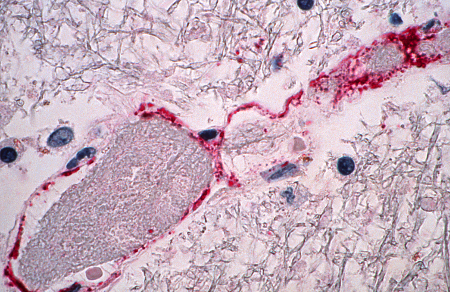 [Figure caption and citation for the preceding image starts]: Photomicrograph showing some of the pathological changes associated with NiV infection affecting the central nervous systemCDC/Brian W.J. Mahy, BSc, MA, PhD, ScD, DSc [Citation ends].
 [Figure caption and citation for the preceding image starts]: Photomicrograph of a central nervous system tissue specimen showing a centrally-located target lesionCDC/Brian W.J. Mahy, BSc, MA, PhD, ScD, DSc [Citation ends].
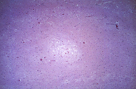
Result
may show medium- to small-sized blood vessel vasculitis with disseminated micro-infarctions; direct neuronal invasion by the virus
Use of this content is subject to our disclaimer