Differentials
Common
Splenic injury
History
history of blunt trauma more common than penetrating trauma; left upper quadrant pain, or referred pain to the left shoulder (Kehr's sign); left lower rib fractures have a high incidence of concurrent splenic injury
Exam
signs of hypovolaemia; left upper quadrant tenderness may be elicited; physical examination is not a sensitive or specific test for diagnosis of splenic injuries
1st investigation
Hepatic injury
History
history of blunt or penetrating trauma; right upper quadrant pain; right lower rib fractures are associated with hepatic injury
Exam
signs of hypovolaemia; may reveal right upper quadrant tenderness or abdominal fullness; physical examination is unreliable
1st investigation
Other investigations
- focused assessment with sonography in trauma (FAST) ultrasound:
intra-abdominal or intracapsular haemorrhage
More - diagnostic peritoneal lavage (DPL):
intra-abdominal haemorrhage
More - hepatic arteriography:
intra-hepatic arterial bleeding
- endoscopic retrograde cholangiopancreatography:
may identify delayed complications of major biliary duct injuries
Renal injury
History
history of blunt or penetrating flank injury; rapid deceleration fall or motor vehicle accident; gross haematuria; pain in abdomen and flank, especially on inspiration
Exam
penetrating wound and/or contusions on flanks or back; fractures of the 11th or 12th ribs; flank tenderness; gross haematuria; pain in abdomen/flank worse with inspiration; costovertebral angle tenderness; haemodynamic instability
1st investigation
Other investigations
Small bowel injury
History
history of penetrating trauma (more common than blunt) leading to peritonitis; often no signs of peritonitis in early period after injury; potentially missed with blunt abdominal trauma where small bowel injury is not suspected
Exam
may be little sign of peritonitis in initial period after injury; later, may have a distended, rigid abdomen with diffuse tenderness; wound penetrating posterior abdominal fascia and/or abdominal wall contusions from blunt trauma or seat belt; potentially missed if stab wound to anterior abdomen misdiagnosed as not having penetrated the posterior abdominal fascia
1st investigation
Other investigations
- diagnostic peritoneal lavage (DPL):
positive if red blood cells >1 X 10¹²/L (>100,000/mm³); >0.5 X 10⁹/L (>500 white blood cells/mm³); presence of bacteria, bile, or food particles
More
Uncommon
Pancreatic injury
History
history of penetrating trauma or localised blunt trauma to upper/mid-abdomen (e.g., handlebar/steering wheel injury); symptoms delayed due to retroperitoneal location of pancreas; vague abdominal pain radiating to back, usually some hours after the traumatic event
Exam
penetrating wound or abdominal contusions, especially on upper/mid-abdomen; signs appear late due to retroperitoneal position; abdominal tenderness, may develop peritoneal irritation with guarding
1st investigation
Other investigations
- magnetic resonance cholangiopancreatography:
ductal injuries, laceration, pseudocyst, or parenchymal injuries
More
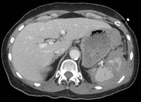
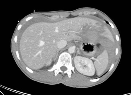
Diaphragmatic injury
History
history of high-velocity blunt abdominal or thoraco-abdominal penetrating trauma; may complain of chest pain, non-specific abdominal pain, or shortness of breath; abdominal pain exacerbated by lying supine
Exam
abdominal contusions and/or penetrating wound, especially if close to costal margin; abdominal pain exacerbated by lying supine; diminished breath sounds on the affected side (left side affected nine times more than right following blunt trauma); auscultation of bowel sounds in lung fields; haemodynamic instability, particularly when lying supine (due to abdominal viscera herniating into thorax and impeding venous return and reducing cardiac output); tachypnoea, tachycardia, shoulder pain, abdominal distension, and/or guarding; missed diaphragmatic injuries associated with abdominal viscera herniation and strangulation
1st investigation
Other investigations
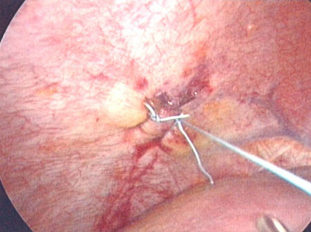
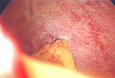
- laparoscopy:
direct visualisation of diaphragmatic injury
More
Stomach injury
History
history of penetrating or blunt abdominal trauma, especially to epigastrium; significant deceleration from fall or traffic accident with full stomach; non-specific abdominal pain
Exam
penetrating traumatic wound and/or contusions consistent with blunt trauma; rapid onset of burning epigastric pain, followed quickly by rigidity and rebound sensitivity; ultimately results in distended, rigid abdomen with diffuse tenderness; potentially missed if stab wound to anterior abdomen misdiagnosed as not having penetrated the posterior abdominal fascia
Other investigations
- nasogastric tube:
blood in nasogastric aspirate
Colorectal injury
History
history of penetrating trauma (more common than blunt) leading to peritonitis; consider colorectal injury in blunt trauma associated with pelvic fractures
Exam
distended, rigid abdomen with diffuse tenderness; gross blood on rectal examination
Other investigations
- CT abdomen/pelvis:
free air under diaphragm or mesenteric haematoma (blunt injuries); contrast extravasation
More
Mesenteric injury
History
history of blunt or penetrating trauma (particularly rapid deceleration or significant force injuries); may be initially asymptomatic or with vague abdominal pain
Exam
abdominal wall ecchymosis; abdominal tenderness with or without peritoneal signs
1st investigation
- CT scan of abdomen:
free intraperitoneal fluid, mesenteric haematoma
More
Other investigations
- diagnostic peritoneal lavage (DPL):
positive if red blood cells >1 X 10¹²/L (>100,000/mm³); >0.5 X10⁹/L (>500 white blood cells/mm³); presence of bacteria, bile, or food particles
More
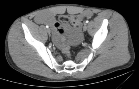
Bladder injury
History
history of blunt or penetrating trauma; associated with pelvic fractures; difficulty voiding and gross haematuria
Exam
lower abdominal tenderness
1st investigation
Other investigations
- CT scan of abdomen and pelvis with intravenous contrast and delayed imaging through pelvis:
free fluid in pelvis
More
Abdominal vascular injury
History
history of penetrating trauma to abdomen or pelvis more common than blunt trauma
Exam
distended abdomen, tachycardia; signs of haemodynamic instability, hypotension; possible loss of pulses to lower extremity
1st investigation
Other investigations
Use of this content is subject to our disclaimer