Investigations
Your Organisational Guidance
ebpracticenet urges you to prioritise the following organisational guidance:
Kleincellige en niet-kleincellige longkanker: diagnose, behandeling en opvolgingPublished by: KCELast published: 2013Cancer du poumon à petites cellules et non à petites cellules : diagnostic, traitement et suiviPublished by: KCELast published: 20131st investigations to order
chest x-ray
Test
A standard posteroanterior chest x-ray is a simple initial step to evaluate cough, chest pain, and/or haemoptysis.
In some centres a lateral chest x-ray may also be performed. A normal chest x-ray does not exclude a diagnosis of lung cancer.
A new abnormality on chest x-ray needs to be further assessed with contrast-enhanced CT.[59] For symptoms such as persistent haemoptysis, it is common practice to omit the chest x-ray and perform CT.
Chest x-rays can be used to track pleural effusions and evaluate for re-expansion of a collapsed lung after an intervention (e.g., endobronchial therapy or radiotherapy).
Result
variable; may detect single or multiple pulmonary nodule(s), mass, pleural effusion, lung collapse, or mediastinal or hilar fullness
contrast-enhanced CT scan of lower neck, thorax, and upper abdomen
Test
Standard and helps to define the primary tumour and evaluate for regional spread.[60]
A new abnormality on chest x-ray needs to be further assessed with contrast-enhanced CT scan.[59] For symptoms such as persistent haemoptysis, it is common practice to omit the chest x-ray and perform CT.
A chest CT should be obtained in patients, especially smokers, with problematic symptoms and a normal chest x-ray. Intravenous contrast is helpful to distinguish lymph nodes from vessels, especially in the hilum.[104]
For mediastinal staging, sensitivity is approximately 60%, specificity is approximately 80%, positive predictive value is between 50% and 55%, and negative predictive value is approximately 85%.[105][106]
CT is useful for evaluation of pleural effusions.[107]
Result
shows size, location and extent of primary tumour; evaluates for hilar and/or mediastinal lymphadenopathy and distant metastases
Investigations to consider
sputum cytology
Test
Specificity is high, but the sensitivity is low.[62][65]
Cytology is more likely to confirm the diagnosis in central lesions than in peripheral lesions.[62][65]
Sputum cytology cannot accurately determine histology or inform appropriate therapy.
Cost may outweigh the usefulness in terms of patient management.
Result
malignant cells in sputum
bronchoscopy
Test
Indicated when CT shows a lesion accessible via bronchoscopy and a biopsy will yield the necessary diagnostic and staging information.
Bronchoscopy, typically performed with a flexible bronchoscope, is an endoscopic procedure in which the proximal bronchial tree can be directly visualised and suspicious areas biopsied.[108] Endobronchial brushings, washings, and alveolar lavage increase the diagnostic yield.
Transbronchial needle aspiration biopsy of accessible parenchymal lesions and mediastinal lymph nodes can be carried out with or without endobronchial ultrasound (EBUS) guidance. The use of EBUS expands the number and levels of mediastinal nodes that can be sampled. Endoscopic ultrasound (EUS) can also be used to access other mediastinal glands plus the left adrenal gland but overall EBUS is a more useful primary procedure.[74]
The sensitivity for centrally located lesions is high (about 90%).[109] The sensitivity for peripheral lesions is lower and depends upon number of biopsies taken, size of mass, and proximity to the bronchial tree. In general, endobronchial biopsy is more sensitive than endobronchial brushings or washings.
Detection of small peripheral lesions (<2 cm) is improved by use of endobronchial ultrasound.[62][110]
Autofluorescent or narrow band imaging can improve the detection of early endobronchial lesions and can be used at the time of initial bronchoscopy or follow-up.[70][71][72]
Result
endobronchial lesions
biopsy
Test
Pathological confirmation of malignancy is the only widely accepted method to make a definitive diagnosis of lung cancer.
Tissue is sampled from bronchoscopy where possible. Transthoracic needle aspiration biopsy, typically using CT guidance, is used to biopsy suspicious peripheral pulmonary lesions that are not accessible with bronchoscopy.
Transbronchial needle aspiration biopsy of accessible parenchymal lesions and mediastinal lymph nodes can be carried out with or without endobronchial ultrasound (EBUS) guidance. The use of EBUS expands the number and levels of mediastinal nodes that can be sampled. Endoscopic ultrasound (EUS) can also be used to access other mediastinal glands plus the left adrenal gland but overall EBUS is a more useful primary procedure.[74]
Lymph node biopsy can provide information about both diagnosis and the stage of disease. Sampling is either of nodes in the supraclavicular fossa or of mediastinal nodes accessed via EBUS, EUS, or surgically via mediastinoscopy, video-assisted thoracoscopic surgery, or an open surgical procedure.[66][67][68][69][79][111]
Samples should provide sufficient tissue to make typing, subtyping, and mutation testing possible. Multiple biopsies or needle aspirations should be taken.
Result
specimen for pathological diagnosis
diagnostic thoracentesis and/or pleural biopsy
Test
For the evaluation of pleural effusions if there is no other evidence of metastatic disease.
Thoracentesis involves placing a needle between the ribs and into the chest to sample fluid that has accumulated in the pleural space.
Thoracentesis may be followed by thoracoscopy, if fluid is negative.
Ultrasound is preferred for all medical pleural procedures and is essential when sampling small pleural effusions.
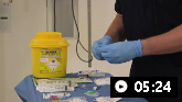
Video demonstrating how to perform a pleural aspiration
Result
presence of malignant cells in sufficient number to allow full subtyping and mutation testing
CT with contrast and/or fluorodeoxyglucose (FDG)-PET
Test
FDG-PET/CT is performed from the skull base to mid-thigh. Positive FDG-PET/CT scan findings for distant disease need pathological or other radiological confirmation. If FDG-PET/CT scan is positive in the mediastinum, lymph node status needs pathological confirmation.[60]
For patients with known metastatic disease, PET is often unnecessary.
PET accuracy for mediastinal staging is as follows: sensitivity between 80% and 85%, specificity approximately 90%, positive predictive value between 80% and 90%, and negative predictive value approximately 93%.[105][112]
In one Cochrane review the accuracy of PET in distinguishing N2/3 disease was found to be variable and dependent on the make of the scanner, subtype of lung cancer, FDG dose, and country of study origin. Thus, it is important for centres to audit the accuracy of their local practice.[113]
The accuracy of PET for diagnosis of lung cancer in pulmonary nodules shows a sensitivity and specificity of 89% and 77%, respectively, but the latter is reduced to 61% in areas with endemic infectious lung disease.[114]
Result
evaluates location and extent of primary tumour; evaluates for hilar and/or mediastinal lymphadenopathy and distant metastases
sampling of the mediastinal lymph nodes: mediastinoscopy and endobronchial ultrasound (EBUS)
Test
Tissue sampling of the mediastinum via mediastinoscopy or endobronchial ultrasound (EBUS) is the most accurate method to stage the mediastinum, and is often indicated following FDG-PET/CT. European guidance recommends combined EBUS and EUS for mediastinal staging of lung cancer; it is less invasive than mediastinoscopy and may avoid unnecessary thoracotomy.[79][80]
Mediastinoscopy is an invasive procedure performed under general anaesthaesia. EBUS is increasingly used instead.
Paratracheal and subcarinal lymph nodes can be assessed with cervical mediastinoscopy.
The overall sensitivity of cervical mediastinoscopy is approximately 80% with a negative predictive value of about 90%.[106]
Result
spread to mediastinal lymph nodes
video-assisted thoracoscopic surgery (VATS)
Test
VATS can be used to evaluate aorticopulmonary window lymph nodes.
VATS also allows access to para-oesophageal and pulmonary ligament lymph nodes.
Result
spread to other intrathoracic lymph nodes
thoracoscopy
Test
Direct visualisation of the pleura using thoracoscopy has a very high degree of specificity for the diagnosis of pleural effusion.
The procedure can be carried out under local anaesthetic, or using video-assisted thoracoscopic techniques under a general anaesthetic.
Result
direct visualisation of pleura; pleural effusion
MRI head
Test
Indicated in patients with early-stage NSCLC if brain metastases are suspected based on symptoms.[60] Patients with locally NSCLC, especially adenocarcinoma, are at higher risk of harbouring brain metastases and should be staged with MRI if potentially curative treatment is proposed.
CT head may be considered if MRI is contraindicated.
Result
brain metastases appear as enhancing parenchymal masses with surrounding oedema
MRI of thoracic inlet
Test
May be helpful in assessing resectability of superior sulcus tumours.[61]
Result
evaluates brachial plexus involvement, invasion of the subclavian vessels, and/or invasion of adjacent vertebral bodies
bone scan
pulmonary function tests (PFT)
Test
All lung cancer patients anticipated to receive chest radiotherapy or surgery should have pulmonary function tests (PFTs) performed, including FEV₁ and diffusing capacity of the lung for carbon monoxide (the latter where spirometry is abnormal or dyspnoea is present).[93][94]
Predicted postoperative lung function should be calculated from baseline values and an estimate of the loss of lung function as a result of surgery.
There is debate on the precise criteria for determining fitness for surgery (and also for potentially curative chemoradiotherapy). Some guidelines suggest using formal cardiopulmonary exercise testing.[94] Other guideline groups suggest the use of risk prediction scores (e.g., Thorascore) in addition to lung function and exercise testing.[61]
Result
spirometry and lung volumes
FBC
Test
Baseline blood counts are necessary before treatment is initiated or invasive procedures are performed.
Chemotherapy, and to a lesser degree radiotherapy, can decrease haematopoiesis, necessitating baseline and periodic analysis of blood counts.
Result
usually normal but may show anaemia
LFTs
Test
May be elevated if hepatic metastases.
Lone elevation of alkaline phosphatase level may indicate bone metastases.
Result
normal or elevated
serum calcium
Test
Elevated in hypercalcaemia of malignancy, more commonly in squamous cell carcinomas.
Result
may be elevated
electrolytes and renal function
Test
Recommended as baseline before treatment is initiated.
Hyponatraemia may be related to the syndrome of inappropriate ADH secretion (SIADH), although this is more commonly seen in small cell lung cancer.
Result
usually normal, although hyponatraemia not uncommon
epidermal growth factor receptor (EGFR) mutation testing
Test
Patients with sensitising EGFR-mutations preferentially benefit from EGFR tyrosine kinase inhibitor therapy over chemotherapy. Testing for mutations of the tyrosine kinase genes that encode EGFR in tumour cells enables targeted therapy to be considered in a subset of patients (ex-light smokers or never-smokers, and those with non-squamous histology, i.e., adenocarcinoma and large cell carcinoma).
Result
positive in some tumours; more prevalent in never-smokers, those with a history of light smoking, and patients with adenocarcinomas; more common in Asian populations
anaplastic lymphoma kinase (ALK) testing
Test
ALK-positive patients preferentially benefit from ALK-inhibitor therapy over chemotherapy. Testing for mutations of the tyrosine kinase genes that encode ALK in tumour cells enables targeted therapy to be considered in a subset of patients (ex-light smokers or never-smokers, and those with non-squamous histology, i.e., adenocarcinoma and large cell carcinoma).
Fusions in the ALK gene can facilitate the proliferation of cancer cells.[115]
Result
positive in some tumours; more prevalent in never-smokers, those with a history of light smoking, and patients with adenocarcinomas
ROS proto-oncogene 1 (ROS1) testing
Test
Testing enables consideration of ROS1-directed therapy with tyrosine kinase inhibitors. ROS1 fusions are usually detected by fluorescence in situ hybridisation (FISH). ROS1 fusions are more prevalent in younger patients, never-smokers, those with a history of light smoking, and patients with adenocarcinomas.[84]
Result
positive in some tumours; more prevalent in younger patients, never-smokers, those with a history of light smoking, and patients with adenocarcinomas
programmed death-ligand 1 (PD-L1) testing
Test
Testing for PD-L1 expression in patients with metastatic non-small cell lung cancer may identify candidates for PD-L1 inhibitor therapy.
PD-L1 status can be evaluated on cytological samples or biopsies; the SP142 assay has lower sensitivity than the 22C3, 28-8, and SP263 assays, which performed similarly in evaluating PD-L1 expression quantity on tumour cells.[91][92]
Result
positive in the majority of tumours
B-Raf proto-oncogene (BRAF) testing
neurotrophin tyrosine receptor kinase (NTRK) fusion testing
Test
Tropomyosin receptor kinase inhibitors are recommended for NTRK fusions.
Result
positive in some tumours
mesenchymal-epithelial transition factor (MET) exon 14 (METex14) skipping mutations
Test
Should be considered. c-MET inhibitors are recommended.
Result
positive in some tumours
re-arranged during transfection (RET) gene mutations testing
KRAS proto-oncogene (KRAS) point mutations testing
Test
Inhibitors of the RAS GTPase family are indicated for the treatment of KRAS G12C-mutated non-small cell lung cancer.
Result
positive in some tumours
ERBB2 (HER2) mutations testing
Test
ERBB2 encodes for HER2, a receptor tyrosine kinase. HER2 overexpression is defined as 3+ tumour cell staining based on IHC. HER2 IHC testing is recommended during disease progression, timed to preserve biopsy tissue. HER2 inhibitor therapy is recommended.
Result
positive in some tumours
neuregulin 1 (NRG1) fusion testing
Test
NRG1 is a HER3 activator, and may induce heterodimerisation with HER2. A HER2/HER3 inhibitor is recommended as subsequent therapy.[89]
Result
positive in some tumours
c-MET overexpression (≥50% immunohistochemistry [IHC] 3+) and EGFR wild-type (no EGFR mutation) testing
Use of this content is subject to our disclaimer