History and exam
Key diagnostic factors
common
malar (butterfly) rash
The characteristic malar or butterfly rash occurs in 30% to 40% of patients, and may be more common in female patients.[50] This erythematous rash extends from the cheeks over the bridge of the nose, sparing the nasolabial folds. Malar rash often recurs after sun exposure.
[Figure caption and citation for the preceding image starts]: Malar rash: butterfly shape, flat, non-tender erythematosus rash over the cheek and noseKumar N et al. BMJ Case Reports. 2013;2013:bcr-2012-008101 [Citation ends].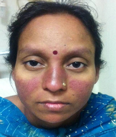 [Figure caption and citation for the preceding image starts]: a) Photograph of a face with skin rashes sparing the bridge of the nose and malar area. b) Photograph of a face showing asymmetric hyperpigmented, polycyclic, and annular scaly plaques with scaling involving pre-auricular area and cheekRajasekharan C et al. BMJ Case Reports. 2013;2013:bcr-2012-007886 [Citation ends].
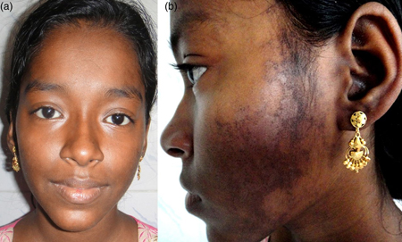
photosensitive rash
Rash occurs after sun exposure. It can be painful and pruritic and usually lasts a few days, healing without scarring. Recent onset of photosensitivity is supportive of the diagnosis.
discoid rash
Erythematous raised patches with adherent keratotic scaling and follicular plugging.
Atrophic scarring may occur in older lesions.
Other diagnostic factors
common
fatigue
A common feature in patients with systemic lupus erythematosus (SLE), occurring in 80% to 100% of patients, but it does not correlate with disease activity.[56][57] Absence of other symptoms suggestive of SLE excludes the diagnosis. The occurrence of fatigue is often independent of signs and symptoms in other systems.
weight loss
Weight loss in people with systemic lupus erythematosus (SLE) may be related to disease activity or its treatment. Patients with SLE may have oesophageal hypomotility leading to dysphagia. Vomiting and diarrhoea may contribute to weight loss. SLE is associated with an increased risk for cancer, and this should be considered as a potential cause of weight loss
fever
Unexplained fever is a common characteristic of systemic lupus erythematosus and is thought to be a presentation of active disease.[1]
oral ulcers
alopecia
Active systemic disease can lead to diffuse patchy alopecia, which is reversible once disease is controlled; however, discoid lesions can leave permanent scarring alopecia.
Usually non-scarring.
Areas of scarring alopecia are more characteristic of chronic discoid lupus.
arthralgia/arthritis
Arthralgia is common in systemic lupus erythematosus (SLE). Inflammatory joint symptoms occur in >50% of patients.[58][62][63]
The arthritis can be similar to rheumatoid arthritis, although classically non-erosive.
Monoarthritis of a large joint is unusual in a patient with SLE and should initially prompt the search for another cause such as infection or avascular necrosis.
Raynaud's phenomenon
Raynaud's phenomenon presents with visible perfusion changes of the hands and feet (vasospasm) in response to cold or emotional stress and should prompt an assessment for other features of systemic lupus erythematosus.[58][62] The nail folds (for capillary nail fold changes) and peripheral pulses should be examined to exclude other causes of Raynaud’s.[58][62]
telangiectasia
Telangiectasia is caused by dilated or broken blood vessels located near the surface of the skin or mucous membrane. They appear as clusters of red or purple lines on the skin.[78]
livedo reticularis
Patients with systemic lupus erythematosus may present with livedo reticularis, a violet web like or rash like pattern under the skin, which is thought to be caused by spasm of the ascending arterioles.[78]
vasculitis
Inflammatory vascular disease can also develop in the form of vasculitis. Vasculitis has been reported in approximately 50% of patients with systemic lupus erythematosus and primarily involves small vessels, medium-sized vessels can be affected; however, large vessel involvement is rare.[78]
chest pain and shortness of breath
Pulmonary manifestations of systemic lupus erythematosus (SLE) include pleuritis, pleural effusions, diffuse interstitial lung disease, pulmonary hypertension and, rarely, pulmonary haemorrhage.[68] Pulmonary embolism should be excluded in patients with SLE presenting with pleuritic chest pain, dyspnoea, and haemoptysis, particularly if antiphospholipid antibodies are positive. Pleural effusions in SLE are usually unilateral and generally exudative. Other causes of a pleural effusion should be excluded.
Shrinking lung syndrome is a rare respiratory manifestation of SLE characterised by dyspnoea, chest pain, a raised hemidiaphragm, and a restrictive pattern on pulmonary function tests.[77]
venous or arterial thrombosis
The presence of antiphospholipid antibodies increases the risk of venous or arterial thrombosis.[92]
hypertension
signs of nephrosis (e.g., oedema)
Renal involvement is present in approximately 50% to 70% of patients, and may be more common in male patients.[50][62] As renal involvement usually develops in the first few years of illness, blood pressure, urinalysis, and estimated glomerular filtration rate (eGFR) should be monitored during regular surveillance.[86][87]
lymphadenopathy
Peripheral lymphadenopathy is more often regional than generalised. The nodes are usually non-tender, vary in size from shotty (clusters of small lymph nodes, each a few mm) to 3-4 cm, and often are in the cervical and axillary regions.
Hilar lymphadenopathy is uncommon.
Patients with lymphadenopathy are more likely to have constitutional manifestations.
Lymphoma and infectious mononucleosis should be excluded.
Histology of lymph node biopsies in systemic lupus erythematosus frequently shows reactive hyperplasia.
abdominal pain, vomiting, or diarrhoea
Systemic lupus erythematosus (SLE) can affect any part of the gastrointestinal tract.[79] Abdominal pain, vomiting, and diarrhoea may be caused by lupus peritonitis or mesenteric artery occlusion, but other causes of an acute abdomen should be excluded.
Although rare, lupus peritonitis may mimic appendicitis. Pancreatitis may be due to SLE, but it is important to exclude treatment such as azathioprine as the underlying cause.
uncommon
nose ulcers
Nose ulcers occur less frequently than oral ulcers.[61] Typically painless but prolonged and recurrent.
poorly localised proximal limb inflammatory pain with weakness
Suggestive of an associated myositis; if present, creatinine phosphokinase will be elevated.
dysrhythmias (e.g., tachycardia), conduction defects, or unexplained cardiomegaly
Myocarditis should be suspected in patients with tachycardia, arrhythmias, conduction defects, or unexplained cardiomegaly.
neuropsychiatric symptoms
Clinical manifestations of neuropsychiatric systemic lupus erythematosus (SLE) may range from subtle cognitive dysfunction to acute confusional states, seizure disorders, and psychosis.[64] Headaches, anxiety, mood, and cognitive disorders are the most frequent neuropsychiatric manifestations of SLE; however, cerebrovascular disease, neuropathies, acute confusional states, and seizure disorders are the most frequent associated with NPSLE.
dysphagia
Dysphagia is less common symptom of the gastrointestinal manifestation of systemic lupus erythematosus, and is due to oesophageal hypomotility.
Risk factors
strong
female sex
The incidence of systemic lupus erythematosus (SLE) is higher in women than in men; reported sex ratios range from 2:1 to 15:1.[6][7][8]
The increased frequency of SLE among women has been associated with the effects of oestrogen.[3][12] Data from the Centres for Disease Control and Prevention National Lupus Registries (2002-2009) revealed that the estimated prevalence of SLE in the US was nine times higher among women (128.7 per 100,000) compared with men (14.6 per 100,000).[9]
Alopecia, photosensitivity, oral ulcers, arthritis, and malar rash may be more common among female patients.[50]
age >30 years
African descent in Europe and US
Data from the US Centres for Disease Control and Prevention National Lupus Registries (2002-2004; 2007-2009) found that prevalence was higher in African-Americans people (230.9 per 100,000 for women and 26.7 per 100,000 for men), than in Hispanic people (120.7 per 100,000 for women and 18.0 per 100,000 for men) or white people (84.7 per 100,000 for women and 8.9 per 100,000 for men).[9]
Reported incidences in Africa are low (0.3 per 100,000 person-years).[3] This may reflect under-diagnosis due to resource shortage.
use of certain drugs
Clinical and serological manifestations can occur in patients taking some drugs.[30][31]
The first reported association was with procainamide, but other commonly implicated drugs include minocycline, terbinafine, sulfasalazine, isoniazid, phenytoin, and carbamazepine.[32][33][34][35][36][37][38][39]
Some of the reported associations between drug use and a subsequent diagnosis of systemic lupus erythematosus may be due to protopathic bias (when a treatment is inadvertently prescribed for an early manifestation of a disease that has not yet been diagnosed).[31]
weak
sun exposure
Exposure to ultraviolet (UV) radiation can exacerbate skin lesions in lupus erythematosus patients (photosensitivity).[25]
Prospective, methodologically robust studies are required to evaluate the relationship between UV-B and incident systemic lupus erythematosus.
family history of SLE
tobacco smoking
air pollution
One meta-analysis of six studies suggests that 6 days of exposure to increased particulate matter 2.5 significantly increases scores on the Systemic Lupus Erythematosus Disease Activity Index.[40]
free crystalline silica
Results from one systematic review and meta-analysis of epidemiological studies support the hypothesis of a possible association between occupational exposure to free crystalline silica and systemic lupus erythematosus, in particular at higher exposure levels, known to induce silicosis.[41]
Use of this content is subject to our disclaimer