Approach
The diagnosis of systemic lupus erythematosus (SLE) can be challenging due to the variable clinical presentation, with non-specific early symptoms and multi-system involvement. It is a clinical diagnosis based on the constellation of signs and symptoms, supported by laboratory test results, after excluding differential diagnoses.[52] Immunological abnormalities are a prominent feature.
Patients may present with clinical features from mild fatigue and fever to severe organ threatening disease.[53] Presentation may include either extrarenal symptoms such as constitutional manifestations, joint manifestations and serositis, mucocutaneous manifestations, neuropsychiatric manifestations, or haematological manifestations, or renal manifestations such as lupus nephritis, or both renal and extrarenal manifestations at the same time.[53] Testing for SLE is guided by patient presentation and clinical experience due to a lack of diagnostic guideline recommendations.
Early diagnosis of SLE (including serological assessment) and regular screening for organ involvement (especially nephritis) are essential to prevent organ damage, improve prognosis, and enhance quality of life.[53][54][55]
History
A patient with suspected SLE may present with a combination of constitutional manifestations, mucocutaneous manifestations, musculoskeletal manifestations, haematological manifestations, cardiopulmonary manifestations, vascular manifestations, gastrointestinal manifestations, or renal manifestations.
Constitutional manifestations
Fatigue, fever, lymphadenopathy, and weight loss are common and occur at some time during the course of the disease.[56][57]
Fatigue occurs in between 80% and 100% of patients, but it does not correlate with disease activity.[56][57] Fatigue from other causes, such as anaemia, hypothyroidism, drugs (e.g., beta-blockers), depression, fibromyalgia, and social stresses, should be considered.
Unexplained fever is a common characteristic of SLE, and is thought to be a presentation of active disease.[1] Exclusion of infection is important before initiating immunosuppressive therapy in a patient with SLE to prevent reactivation or exacerbation of chronic infection.[58] Fever persisting despite treatment with a non-steroidal anti-inflammatory drug (NSAID) or paracetamol should raise suspicion for an infectious or drug-related aetiology. All patients presenting with persistent fever should have an appropriate symptom-targeted infection screen.
Weight loss in people with SLE may be related to disease activity or its treatment. Patients with SLE may have oesophageal hypomotility leading to dysphagia. Vomiting and diarrhoea may contribute to weight loss. SLE is associated with an increased risk for cancer, and this should be considered as a potential cause of weight loss.
Peripheral lymphadenopathy is more often regional than generalised. The nodes are usually non-tender, vary in size from shotty (clusters of small lymph nodes, each a few mm) to 3-4 cm, and are often in the cervical and axillary regions. Hilar lymphadenopathy is uncommon. Patients with lymphadenopathy are more likely to have constitutional manifestations. Lymphoma and infectious mononucleosis should be excluded. Histology of lymph node biopsies in SLE frequently shows reactive hyperplasia.
Mucocutaneous manifestations
Cutaneous manifestations of SLE are frequently seen; the characteristic malar or butterfly rash occurs in 30% to 40% of patients, and may be more common in female patients.[50] This erythematous rash extends from the cheeks over the bridge of the nose, sparing the nasolabial folds. It can be painful and pruritic, usually lasts for a few days, and heals without scarring. Malar rash often recurs after sun exposure. When rash occurs both above and below the neck it is referred to as acute generalised cutaneous lupus rash. Recent onset of photosensitivity is supportive of the diagnosis.
[Figure caption and citation for the preceding image starts]: Malar rash: butterfly shape, flat, non-tender erythematosus rash over the cheek and noseKumar N et al. BMJ Case Reports. 2013;2013:bcr-2012-008101 [Citation ends].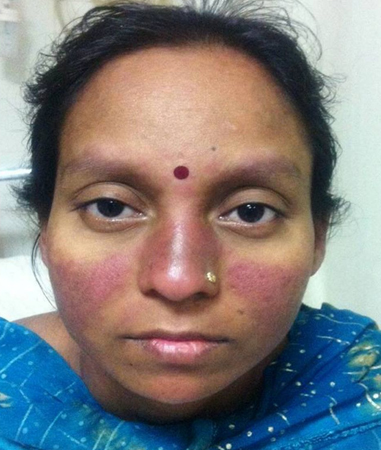
[Figure caption and citation for the preceding image starts]: a) Photograph of a face with skin rashes sparing the bridge of the nose and malar area. b) Photograph of a face showing asymmetric hyperpigmented, polycyclic, and annular scaly plaques with scaling involving pre-auricular area and cheekRajasekharan C et al. BMJ Case Reports. 2013;2013:bcr-2012-007886 [Citation ends].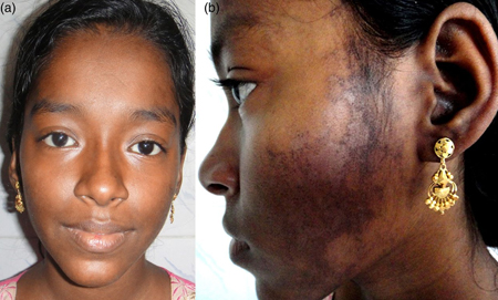
Other distinct categories of rash include discoid lupus, which presents as erythematous raised patches with adherent keratotic scaling and follicular plugging. Atrophic scarring may occur in older lesions. The latter patterns are less likely to be associated with systemic disease, but many patients are antinuclear antibody (ANA)-positive.
The most frequently associated oral manifestations with SLE are oral ulcers, hyposalivation, pigmentations, glossodynia, cleft tongue, cheilitis, arthritis, and secondary Sjögren's syndrome.[59] Oral mucosal lesions have been reported in up to 31% of patients with SLE, with oral ulcers in 30%.[60]Nose ulcers are less common.[61] Both oral and nose ulcers are usually large and painless, in contrast to herpetic lesions.[61] These ulcers often improve with simple local measures and their course parallels the disease course.
Active systemic disease can lead to diffuse patchy alopecia, which is reversible once disease is controlled; however, discoid lesions can leave permanent scarring alopecia.
Musculoskeletal manifestations
Musculoskeletal symptoms are common, occurring in the majority of patients at some time during the course of the illness.[58][62][63] Determining the distribution and the nature of the symptoms is helpful. A diurnal variation in pain, worse in the mornings with associated stiffness, suggests an underlying inflammatory component. Arthritis tends to be symmetrical and is typically non-erosive. Although uncommon, joint deformity may occur; joint deformity in the absence of erosive disease is referred to as Jaccoud's arthritis. Correctable ulnar deviation and joint subluxations in the hands in the absence of radiological damage are characteristic. Patients with SLE may also develop myositis leading to muscle weakness and pain. Septic arthritis should always be excluded in a patient presenting with monoarthritis. The affected joint should be aspirated and fluid sent for microscopy and culture.
Diffuse musculoskeletal pain without a distinct diurnal variation may suggest co-existing fibromyalgia. See Fibromyalgia.
Neuropsychiatric manifestations
Neuropsychiatric manifestations of SLE (NSPLE) are diagnostically challenging. They may range from subtle cognitive dysfunction to acute confusional states, seizure disorders, and psychosis.[64] Headaches, anxiety, mood, and cognitive disorders are the most frequent neuropsychiatric manifestations of SLE; however, cerebrovascular disease, neuropathies, acute confusional states, and seizure disorders are frequently associated with NPSLE, suggesting more than one underlying causative factor.[64]
The American College of Rheumatology has proposed a set of definitions for 19 neuropsychiatric syndromes, hoping to homogenise the terminology for clinical practice.[65] The syndromes are split into central and peripheral nervous system:[65]
Central
Aseptic meningitis
Cerebrovascular disease
Demyelinating syndrome
Headache (including migraine and benign intracranial hypertension)
Movement disorder (chorea)
Myelopathy
Seizure disorders
Acute confusional state
Anxiety disorder cognitive dysfunction
Mood disorder
Psychosis
Peripheral
Acute inflammatory demyelinating polyradiculoneuropathy (Guillain-Barr´e syndrome)
Autonomic disorder
Simple/multiple mononeuritis
Myasthenia gravis
Cranial neuropathy
Plexopathy
Polyneuropathy
Additionally, these syndromes are classified into focal neurological syndromes and diffuse neuropsychiatric syndromes:[65]
Neurological syndromes - focal
Seizure disorder
Aseptic meningitis
Demyelinating syndrome
Myelopathy
Headache (including migraine and benign intracranial hypertension)
Cerebrovascular disease
Movement disorders
Autonomic disorders
Myasthenia gravis
Polyneuropathy
Cranial neuropathy
Plexopathy
Neuropsychiatric syndromes - diffuse
Anxiety disorders
Psychosis
Acute confusional state
Cognitive dysfunction
Mood disorders
Two pathological mechanisms have been demonstrated to contribute to neuropsychiatric SLE:[64]
Autoimmune or inflammatory: due to inflammatory mediators or autoantibodies with either intrathecal immune complex formation or disrupted blood-brain barrier.
Ischaemic or thrombotic: leading to cerebral microangiopathy, vascular occlusion, and haemorrhage. Accelerated atherosclerosis, immune complex deposition, and immune-mediated vascular injury interplay in this pathway.
Haematologic manifestations
Haematologic manifestations are common in SLE at the time of diagnosis. The most frequent include anaemia, leukopenia, lymphopenia, thrombocytopenia, lymphadenopathy, and splenomegaly, which may be related to SLE disease activity and/or caused by immunosuppressive treatment.[66]
Anaemia is usually secondary to chronic disease and improves once control of disease activity has been established. Haemolytic anaemia occurs in approximately 10% of patients with SLE and can be very severe.[66]
Leukopenia is usually due to lymphopenia, and to a lesser extent neutropenia. Thrombocytopenia is also frequently seen and other causes should be excluded. The presence of antiphospholipid antibodies increases the risk of venous and arterial thromboses.
Cardiopulmonary manifestations
SLE may present with diverse cardiopulmonary manifestations including pericarditis, myocarditis, valvular disease, atherosclerosis, thrombosis, arrhythmias, pleuritis, pleural effusions, diffuse interstitial lung disease, pulmonary hypertension and, rarely, pulmonary haemorrhage.[67][68] Pulmonary embolism should be excluded in patients with SLE presenting with pleuritic chest pain, dyspnoea, and haemoptysis, particularly if antiphospholipid antibodies are positive. Pleural effusions in SLE are usually unilateral and generally exudative. Other causes of a pleural effusion should be excluded. Pleuritis and pericarditis are more common than peritonitis in patients with SLE.[69][70] Patients may also develop hypertension as part of cardiopulmonary manifestations.
The risk of cardiovascular events (myocardial infarction and stroke) is two- to threefold higher in patients with SLE compared with the general population.[71][72][73][74][75]
Myocarditis should be suspected in patients with tachycardia, arrhythmias, conduction defects, or unexplained cardiomegaly. One systematic review and meta-analysis demonstrated that patients with SLE have a lower speckle tracking echocardiography compared with people without SLE; this suggests the presence of an impaired myocardial function involving both the left and right ventricle.[76]
Non-bacterial Libman-Sacks endocarditis is uncommon but may be a consequence of a concurrent antiphospholipid syndrome, and should be excluded in case of new murmurs in the presence of antiphospholipid antibodies.
Shrinking lung syndrome is a rare respiratory manifestation of SLE characterised by dyspnoea, chest pain, a raised hemidiaphragm, and a restrictive pattern on pulmonary function tests.[77]
Vascular manifestations
Extracardiac vascular manifestations of SLE include telangiectasia, vasculitis, livedo reticularis, Raynaud's phenomena, and venous or arterial thromboembolism; all of which may occur either alone or in different combinations.[78] Telangiectasia is caused by dilated or broken blood vessels located near the surface of the skin or mucous membrane. They appear as clusters of red or purple lines on the skin. Patients with SLE may present with livedo reticularis, a violet web like or rash like pattern under the skin, which is thought to be caused by spasm of the ascending arterioles.
Inflammatory vascular disease can also develop in the form of vasculitis. Vasculitis has been reported in approximately 50% of patients with SLE and primarily involves small vessels, medium-sized vessels can be affected; however, large vessel involvement is rare.[78]
Raynaud's phenomenon presents with visible perfusion changes of the hands and feet (vasospasm) in response to cold or emotional stress, and should prompt an assessment for other features of SLE. The nail folds (for capillary nail fold changes) and peripheral pulses should be examined.[58][62] The skin over the dorsum of the hands should be checked for sclerodactyly, and features of systemic sclerosis or mixed connective tissue disease should be considered.
The presence of venous or arterial thromboembolism may suggest the possibility of antiphospholipid antibody syndrome.
Gastrointestinal manifestations
SLE can affect any part of the gastrointestinal tract.[79] Abdominal pain, vomiting, and diarrhoea may be caused by lupus peritonitis or mesenteric artery occlusion, but other causes of an acute abdomen should be excluded. Dysphagia is less common and is due to oesophageal hypomotility.
Although rare, lupus peritonitis may mimic appendicitis. Pancreatitis may be due to SLE, but it is important to exclude treatment such as azathioprine as the underlying cause. Chronic active hepatitis may occur in SLE.
Renal manifestations
Renal involvement is present in approximately 50% to 70% of patients, and may be more common in male patients.[50][62] Lupus nephritis is more common in Hispanic and black people, and those with more severe disease in other organ systems. People with antibodies to double-stranded (ds)DNA are more likely to develop glomerulonephritis. Most people with glomerulonephritis are asymptomatic; other presentations include hypertension, nephrotic syndrome, or renal failure. Urinalysis may demonstrate the presence of haematuria, casts (red cell, granular, tubular, or mixed), or proteinuria.
Initial tests for all patients
If a patient presents with a combination of symptoms and manifestations that could indicate SLE, and other diagnoses have been excluded, SLE should be suspected. The following tests should be performed in all patients with suspected SLE:
Full blood count with differential, and clotting screen
A prolongation of the partial thromboplastin time suggests the presence of lupus anticoagulant and should prompt checking of antiphospholipid antibodies.
Infection screen
The infection screen should include blood and urine cultures in febrile patients. Septic arthritis should always be excluded in a patient presenting with a mono-arthritis as it needs to be treated expeditiously
Urea and electrolytes
To exclude or confirm possible renal involvement
Erythrocyte sedimentation rate (ESR) and C-reactive protein (CRP)
Elevated ESR and CRP are suggestive of active disease, but infection must be excluded
Urinalysis
Urinalysis is indicated in all patients with suspected SLE, and regularly in patients with SLE (even in the absence of symptoms).
Antinuclear antibody (ANA) test (anti-double stranded DNA antibody, or anti-Smith antibody)
The American College of Rheumatology recommends the immunofluorescence ANA test using human epithelial type 2 (HEp-2) substrate for ANA testing.[80][81] A positive ANA alone is not diagnostic of SLE because it may be positive in other connective tissue diseases such as rheumatoid arthritis, systemic sclerosis, Sjogren's syndrome, thyroid disease, chronic infectious diseases, and inflammatory bowel disease, and in patients treated with certain drugs such as procainamide, hydralazine, isoniazid, and chlorpromazine. A low, or high (although less common), titre ANA can occur in healthy people.[82] Therefore, a positive ANA has to be interpreted in the light of the clinical history and symptoms.
ANA can be negative in SLE, especially in anti-Ro-antibody-positive lupus (Ro is also known as Sjogren's syndrome A or Sjogren's antibody).
Anti-dsDNA and anti-Smith antibodies are highly specific for SLE and often are confirmatory of the diagnosis, if present.[83][84] High titres of anti-dsDNA antibodies are markers of disease activity and high levels predict worse outcome in lupus nephritis.
Additional tests according to specific manifestations
Haematological manifestations
Coombs' test should be ordered if initial blood count shows anaemia and features of haemolysis, such as elevated MCV and reticulocyte count.
Complement levels can be used in the setting of significant organ manifestations such as cerebritis or nephritis. Sequential rather than single measurements are necessary to be of value, in order to follow response to treatment or confirm worsening disease.
Vascular manifestations
Antiphospholipid antibodies should be ordered in patients with SLE, and thereafter in those with an adverse pregnancy history or arterial/venous thrombotic events.[85]
Mucocutaneous manifestations
Skin biopsy is often not necessary to confirm the diagnosis of mucocutaneous manifestations as these are typically diagnosed clinically, but would be performed if diagnosis is in doubt. Skin biopsy of affected areas may show classic immune deposits at the dermal-epidermal junction on immunofluorescence or non-specific inflammation.
Musculoskeletal manifestations
If there are indications of musculoskeletal involvement, x-rays of affected joints should be requested.
If there is evidence of poorly localised proximal limb inflammatory pain with weakness, creatinine phosphokinase may be done to exclude myositis.
Renal manifestations
As renal involvement usually develops in the first few years of illness, blood pressure, urinalysis, and estimated glomerular filtration rate (eGFR) should be monitored during regular surveillance (every 6-12 months), or when there is suspicion of a disease flare.[86] The testing panel should include:[87]
Urinalysis (dipstick and sediment)
Spot protein-creatinine ratio
Serology (anti-dsDNA and complement)
24-hour urine collection for protein or spot urine for protein/creatinine ratio should be performed if the urinalysis is abnormal, defined as:[87]
Abnormal proteinuria assessed by dipstick protein ≥2+ (any level of specific gravity)
Dipstick protein 1+ (low specific gravity)
Spot PCR >500 mg/g (50 mg/mmol)
Urine sediment positive for acanthocytes (≥5%), red blood cell casts or white blood cell casts
A renal ultrasound should be performed in patients with abnormal urinary sediment to exclude other causes of renal impairment.
A renal biopsy is the most sensitive and specific test for confirming the diagnosis of lupus nephritis and grading the extent of involvement by the International Society of Nephrology (ISN) and Renal Pathology Society (RPS) classification of lupus nephritis.[2] If there is evidence of decreased or decreasing GFR, that is, eGFR that is below the expected level based on age and clinical history, or decreasing eGFR with no attributable cause other than SLE, after a repeat test to confirm the initial findings, a kidney biopsy should be considered.[86][87]
Neuropsychiatric manifestations
Neuropsychiatric manifestations are typically diagnosed clinically with neurological and psychiatric evaluation in addition to the evaluation of general SLE activity, cardiovascular risk factors, atherosclerotic disease, and thrombotic events.[64] Brain MRI may be necessary if the diagnosis is in doubt and will show small focal areas of increased signal, which could be areas of inflammation. These lesions may resolve with treatment.
In patients with SLE with progressive cognitive loss, clinical evidence of SLE activity should be sought, and other causes (such as infection, electrolyte disturbance, vitamin or thyroid deficiency, or medication side effects) need to be excluded.
Anti-ribosomal P is significantly associated with neuropsychiatric manifestations of SLE, including CNS involvement, depression, and psychosis.[88][89] Standardisation of anti-ribosomal P assays is required.
Cardiopulmonary manifestations
Patients presenting with cardiopulmonary symptoms should have a chest x-ray and ECG routinely.[90] Depending on the presenting complaint, the following tests may be needed:
Echocardiogram
Pulmonary function tests
Chest CT
Patients presenting with pleural effusions need pleural aspiration to confirm the cause.
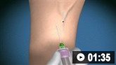
How to take a venous blood sample from the antecubital fossa using a vacuum needle.
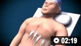
How to record an ECG. Demonstrates placement of chest and limb electrodes.
Use of this content is subject to our disclaimer