Images and videos
Images


Mallory-Weiss tear
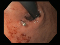
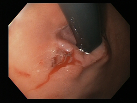
Mallory Weiss tear after application of through-the-scope clip results in haemostasis
From the personal collection of Douglas Adler; used with permission
See this image in context in the following section/s:
Mallory-Weiss tear
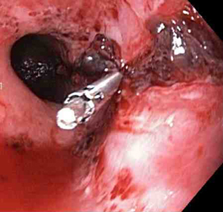
A through-the-scope clip deployed in the centre of the lesion (no previous adrenaline was infused in this case)
From the collection of Juan Carlos Munoz, MD, University of Florida
See this image in context in the following section/s:
Mallory-Weiss tear
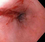
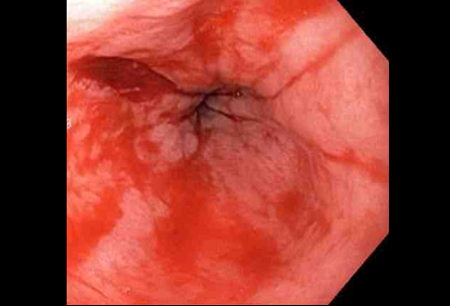
Bleeding Mallory Weiss Tear viewed on retroflexion
From the personal collection of Douglas Adler; used with permission
See this image in context in the following section/s:
Mallory-Weiss tear
Adrenaline is injected locally around the site of the Mallory-Weiss tear
From the collection of Juan Carlos Munoz, MD, University of Florida
See this image in context in the following section/s:
Mallory-Weiss tear
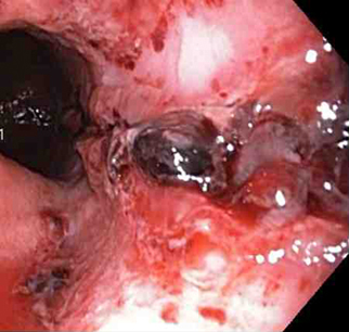
Non-bleeding adherent clot
From the collection of Juan Carlos Munoz, MD, University of Florida
See this image in context in the following section/s:

Mallory-Weiss tear
Mallory-Weiss tear after adrenaline injection (the bleeding has stopped, allowing better visualisation of the lesion)
From the collection of Juan Carlos Munoz, MD, University of Florida
See this image in context in the following section/s:
Mallory-Weiss tear
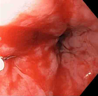
Actively bleeding tear appears as a red longitudinal defect with normal surrounding mucosa
From the collection of Juan Carlos Munoz, MD, University of Florida
See this image in context in the following section/s:

Mallory-Weiss tear
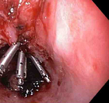
Three haemoclips deployed to complete closure of the mucosal defect
From the collection of Juan Carlos Munoz, MD, University of Florida
See this image in context in the following section/s:
Videos
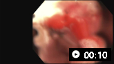 Bleeding Mallory Weiss tear
From the personal collection of Douglas Adler; used with permission
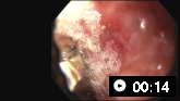 Mallory Weiss tear following cauterisation with a bipolar probe
From the personal collection of Douglas Adler; used with permission
Use of this content is subject to our disclaimer